

Material Characterization
Device Characterization
Material Characterization
Device Characterization
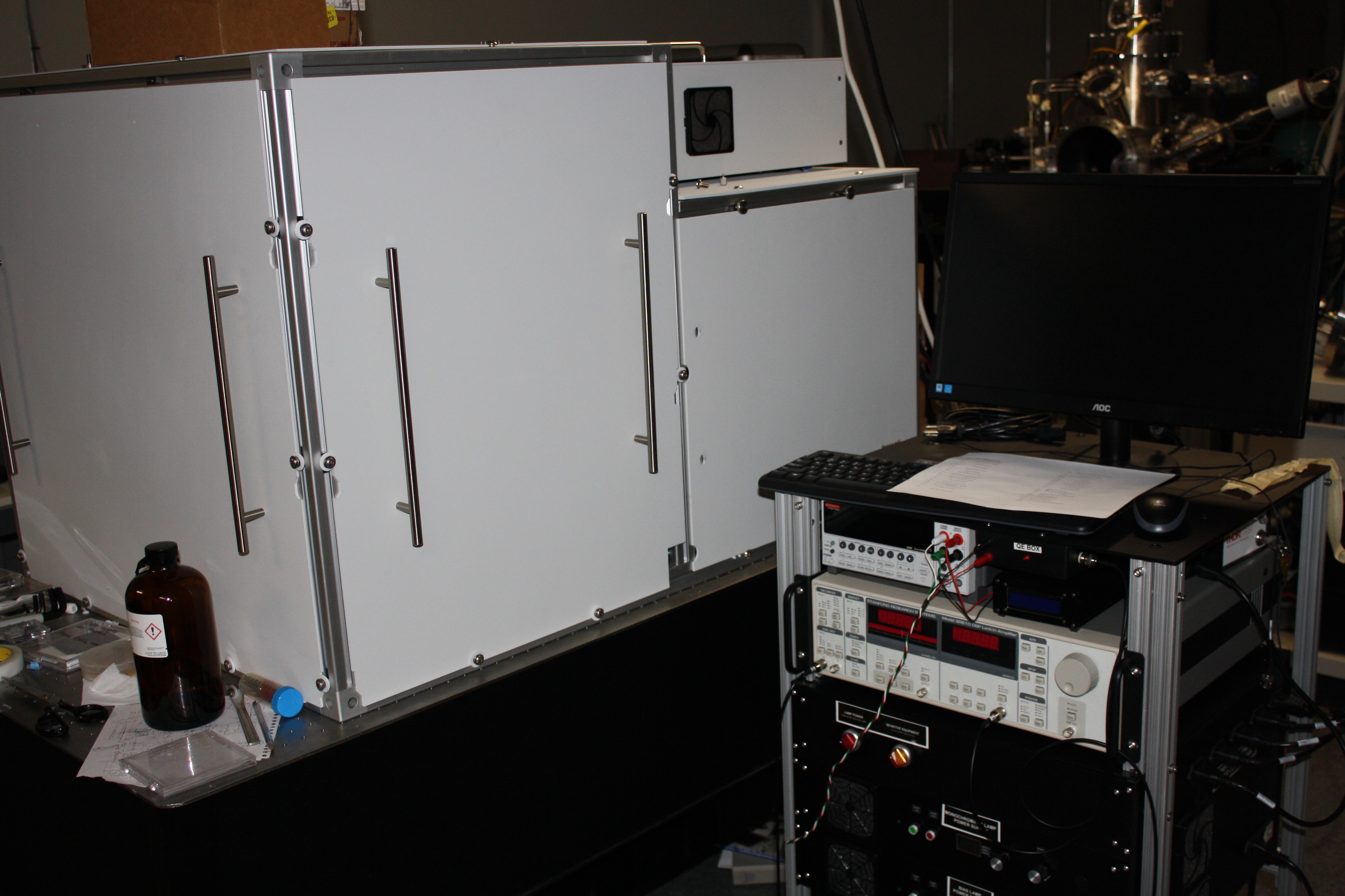
| External Quantum Efficiency System | |
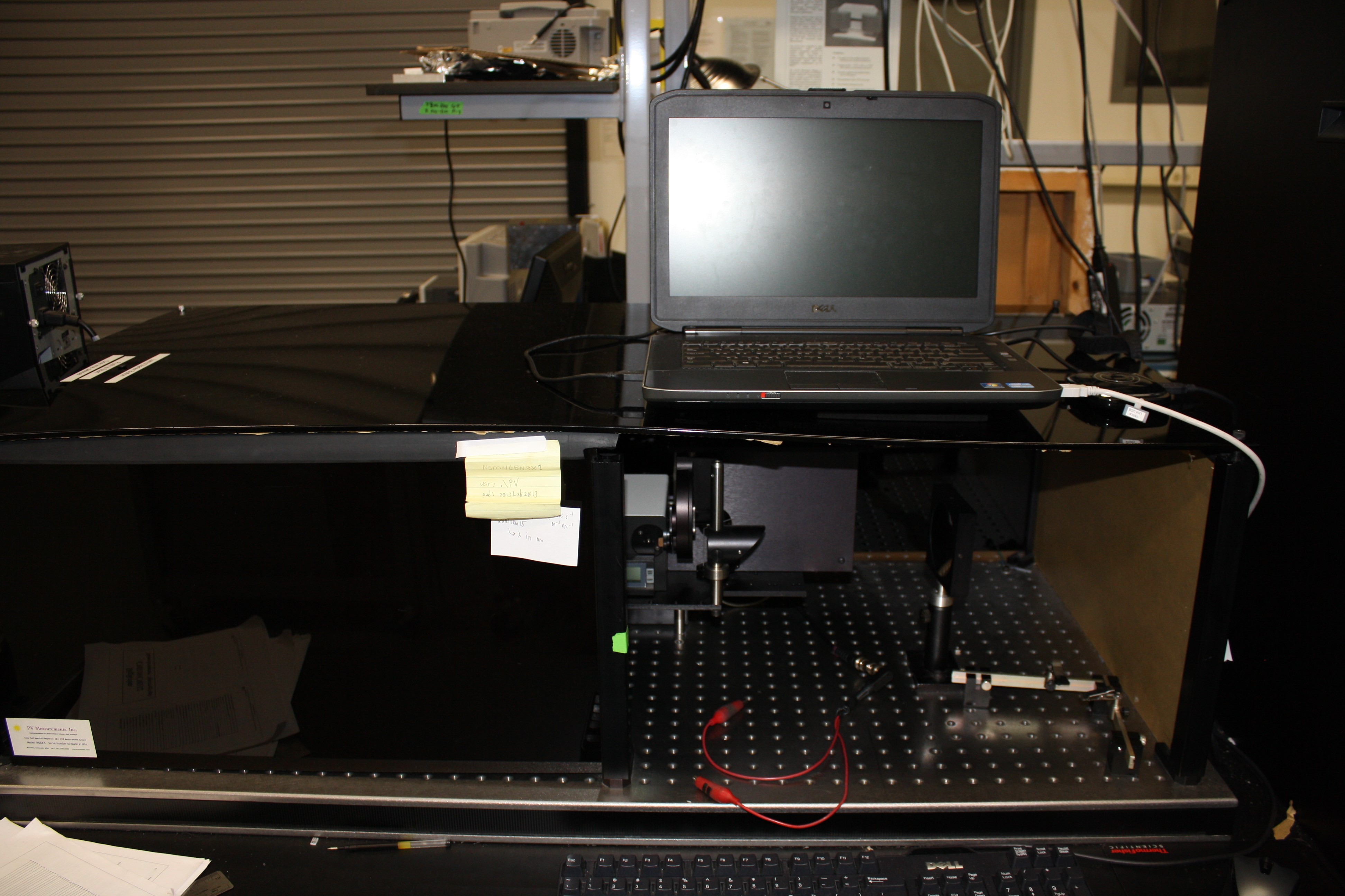 |
Our external quantum efficiency (EQE) system consists of a monochrometer, filter wheel, and reference monitor cell to allow for accurate quantum efficiency measurements. The wavelength range of this system is 300 nm to 1100 nm. This system was built by PV Measurements, and the software was modified in-house. |
| JVT System | |
 |
Our JVT system utilizes a closed loop cryostat to get to temperatures as low as 10 K. The JV response of the samples can be measured under both dark and illuminated conditions during the same temperature scan. |
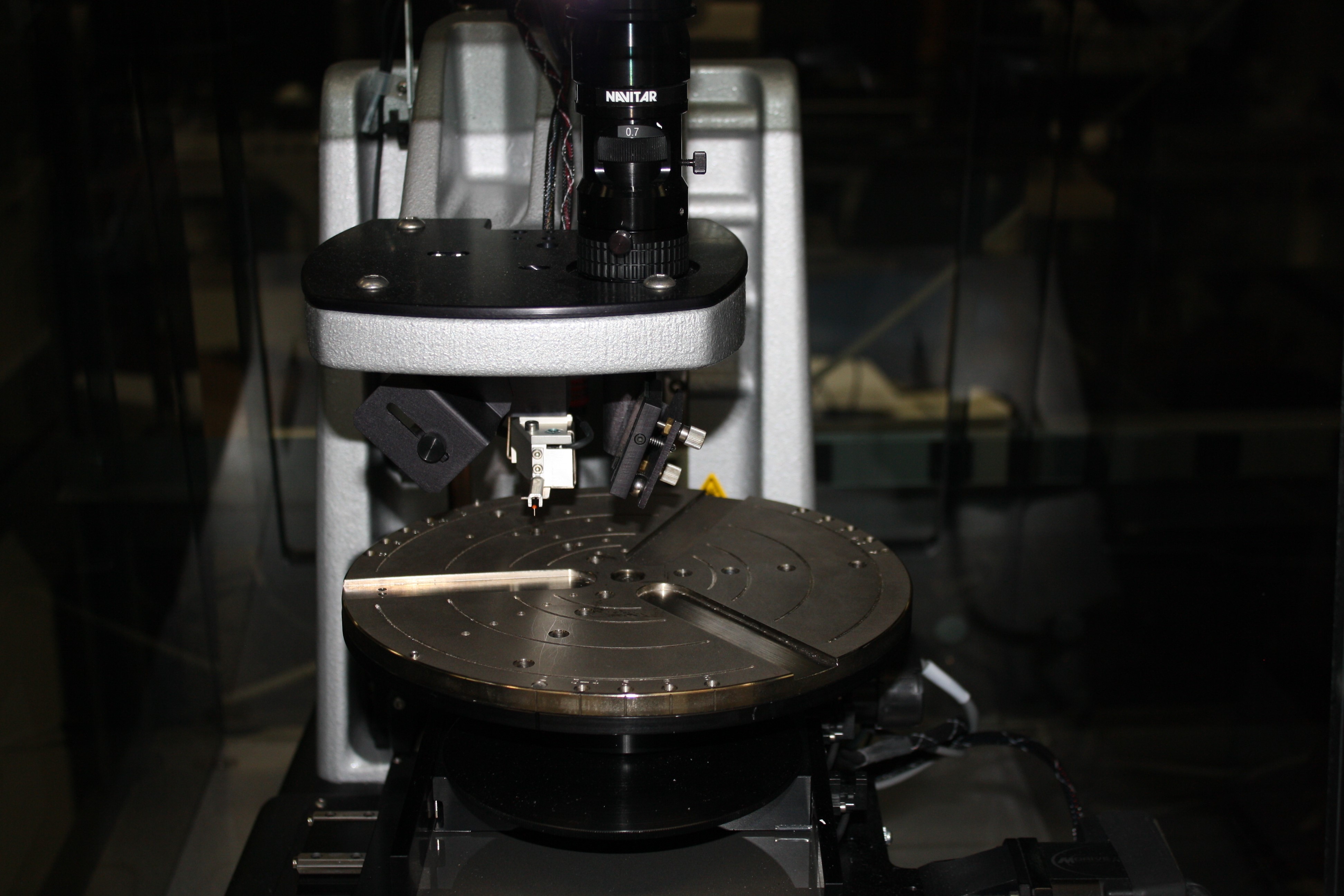
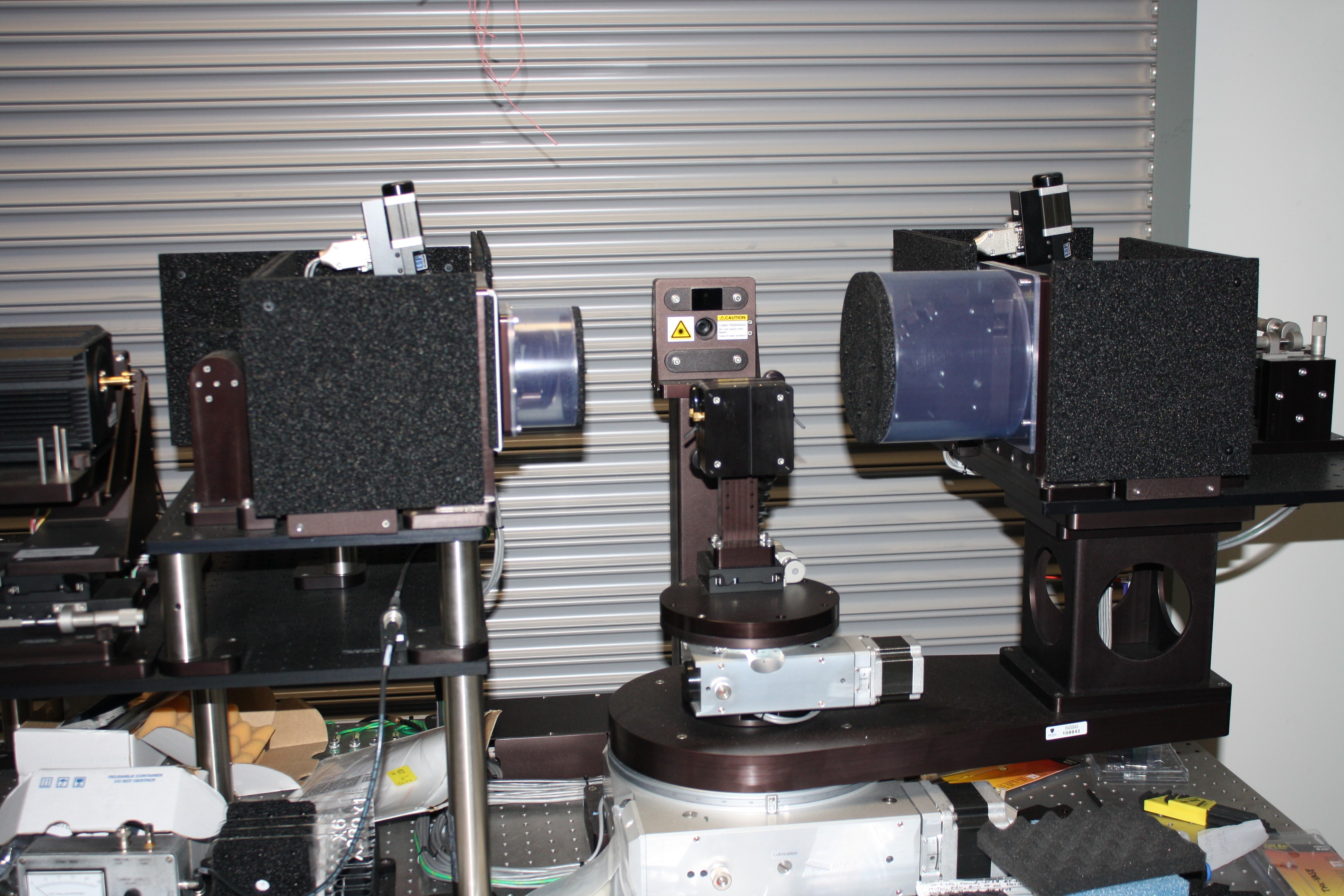
| Laser Beam Induced Current Measurement System | |
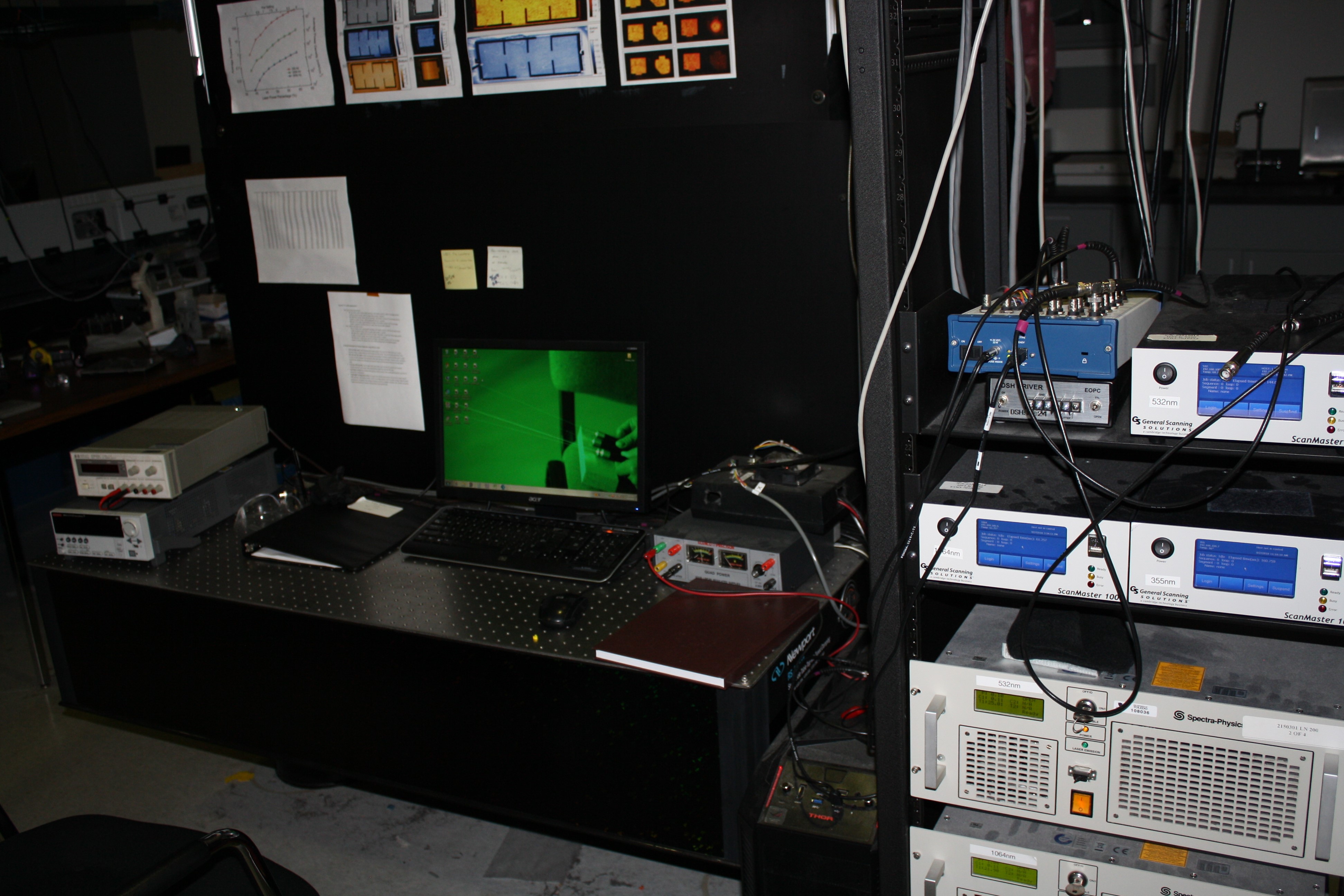 |
Our LBIC system can measure small area samples up to full panel size samples with resolution of 40 microns. Three laser wavelengths (1064 nm, 532 nm, and 355 nm) are available, as is a bias light for spatially characterizing tandem devices. We have also developed an environmental chamber to measure device performance during while controlling the ambient environment. This system was built in-house and is fully automated. |
| Solar Flash Table/JV System | |
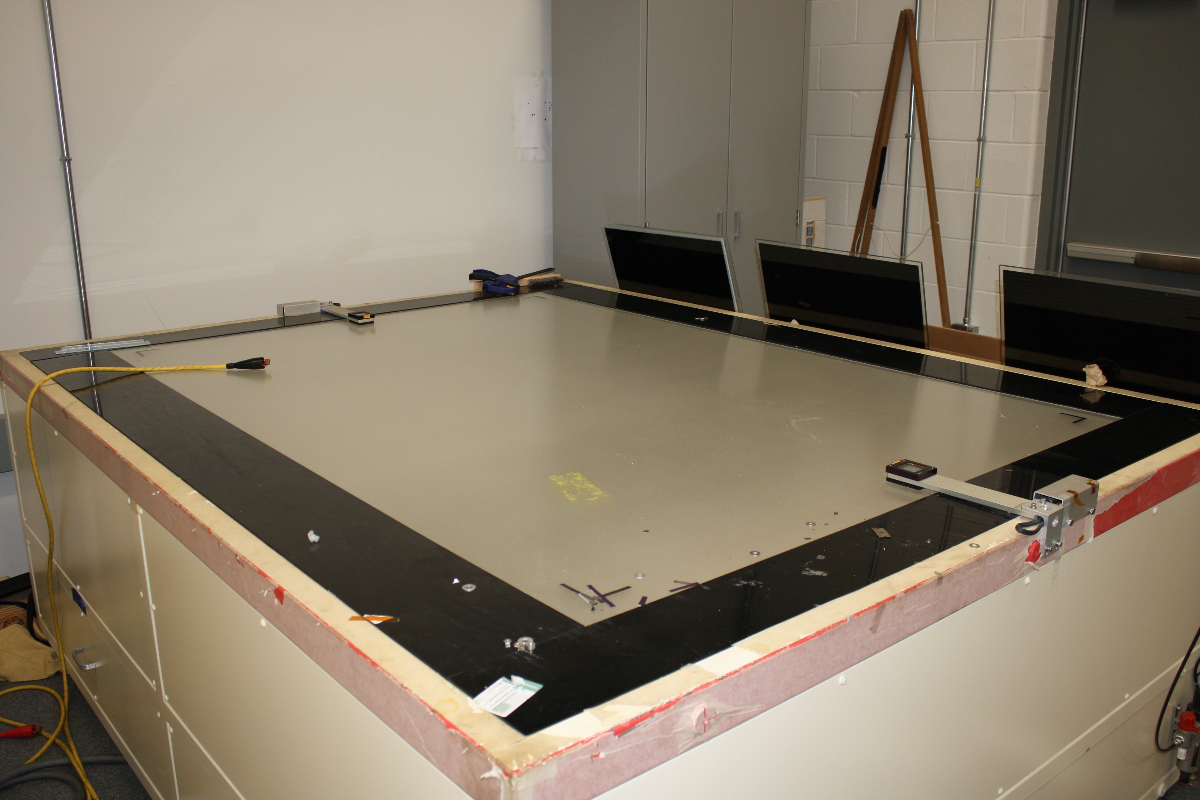 |
Our solar flash table/JV system is able to test large photovoltaic panels by illumination approximating natural sunlight. This type solar simulator is the flashed simulator which uses flash tubes with typical durations of several milliseconds. This system is made by Spire. |
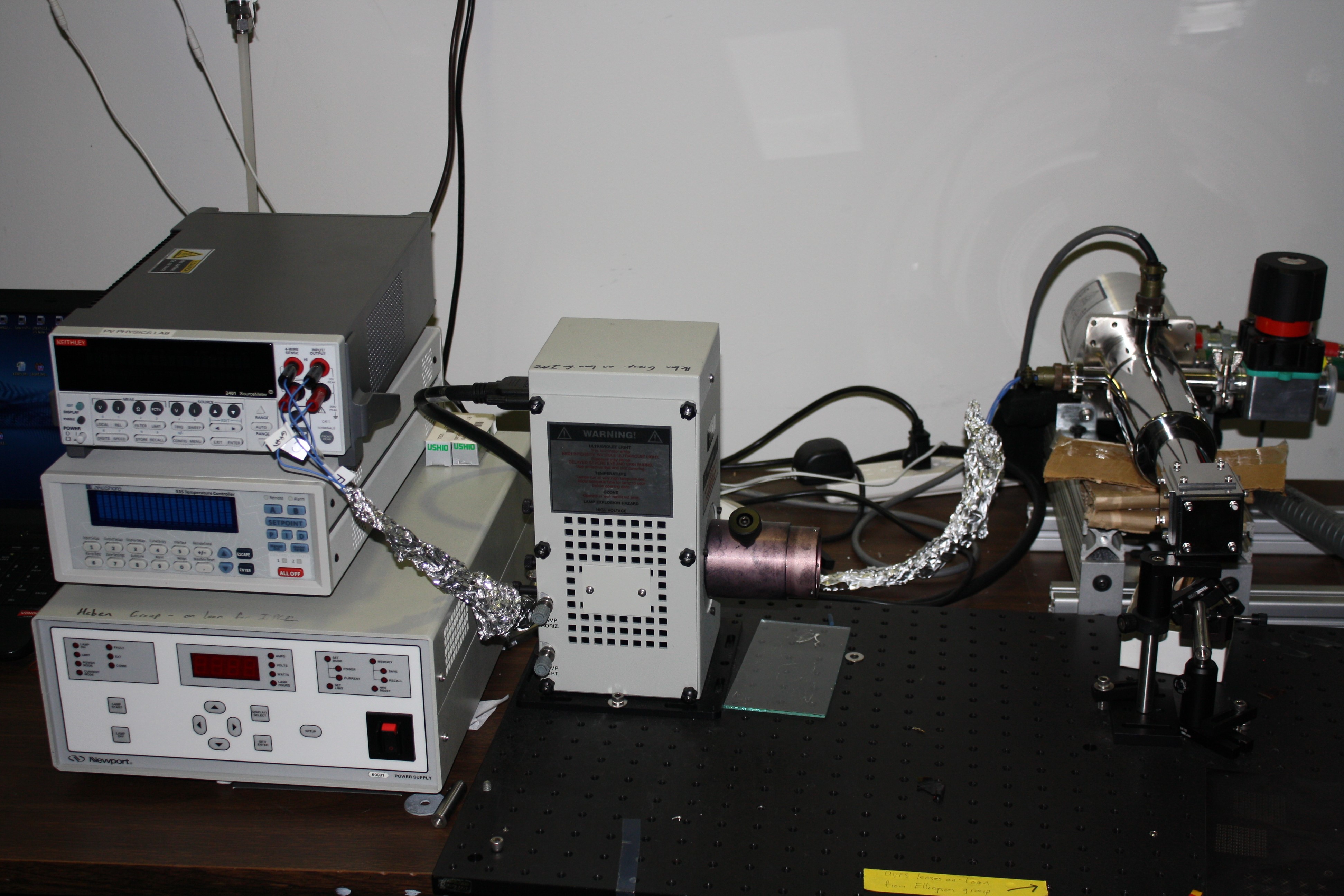
| Solar Simulator JV System | |
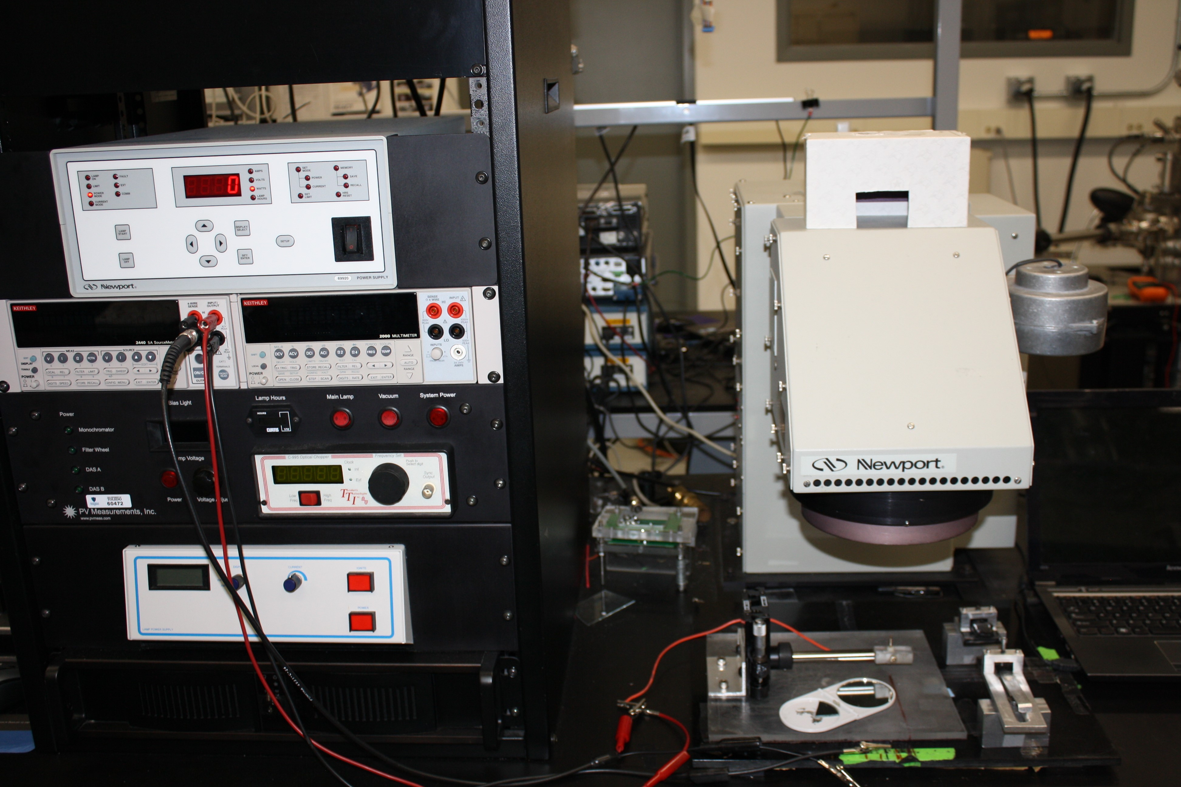 |
Our solar simulator is a Class A Solar Simulator that uses a xenon arc lamp and has 4 in. x 4 in. uniformity. A source meter is used to measure the performance of the cell. The system is capable of measuring substrate and superstrate configuration devices. A multiplexing system and environmental chamber is available. This system was made by PV Measurements, and the software was updated in-house. |
Material Charaterization
| Atomic Force Microscope | |
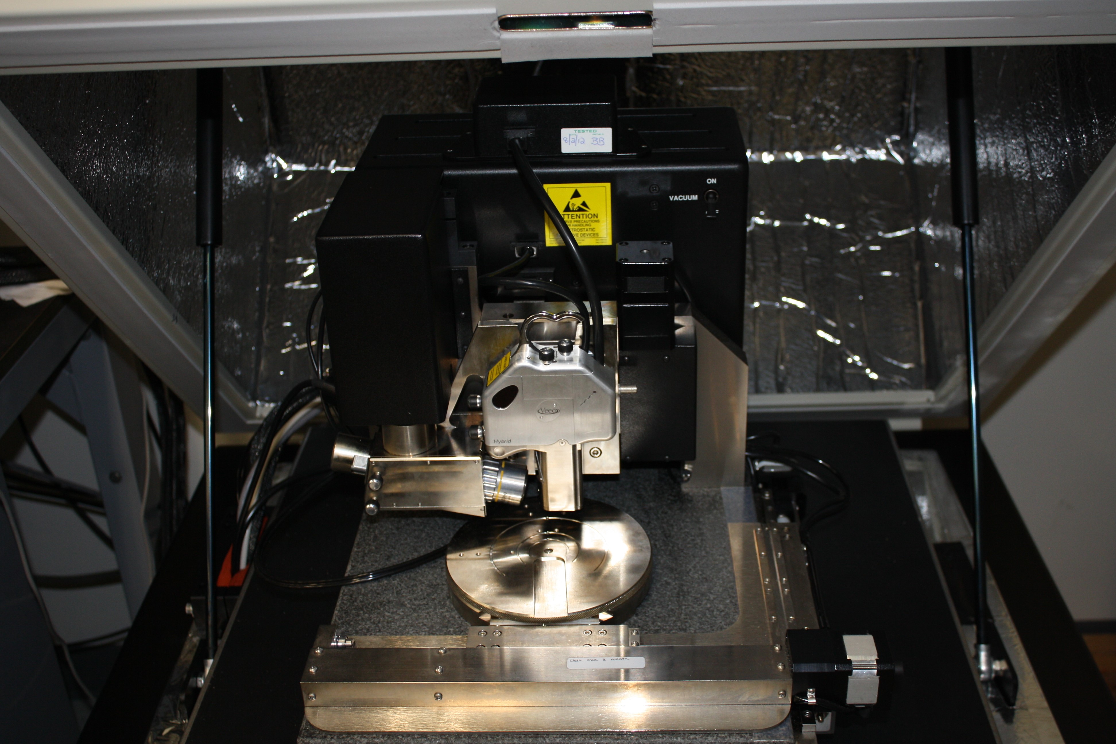 |
Our atomic force microscope (AFM) is a very high-resolution type of scanning probe microscopy, with demonstrated resolution on the order of fractions of a nanometer. The system is configured for contact and non-contact mode. The samples, up to 6 in. in diameter, is held in place using a vacuum chuck. The system was made by Veeco. |
| Auger Electron Spectrometer | |
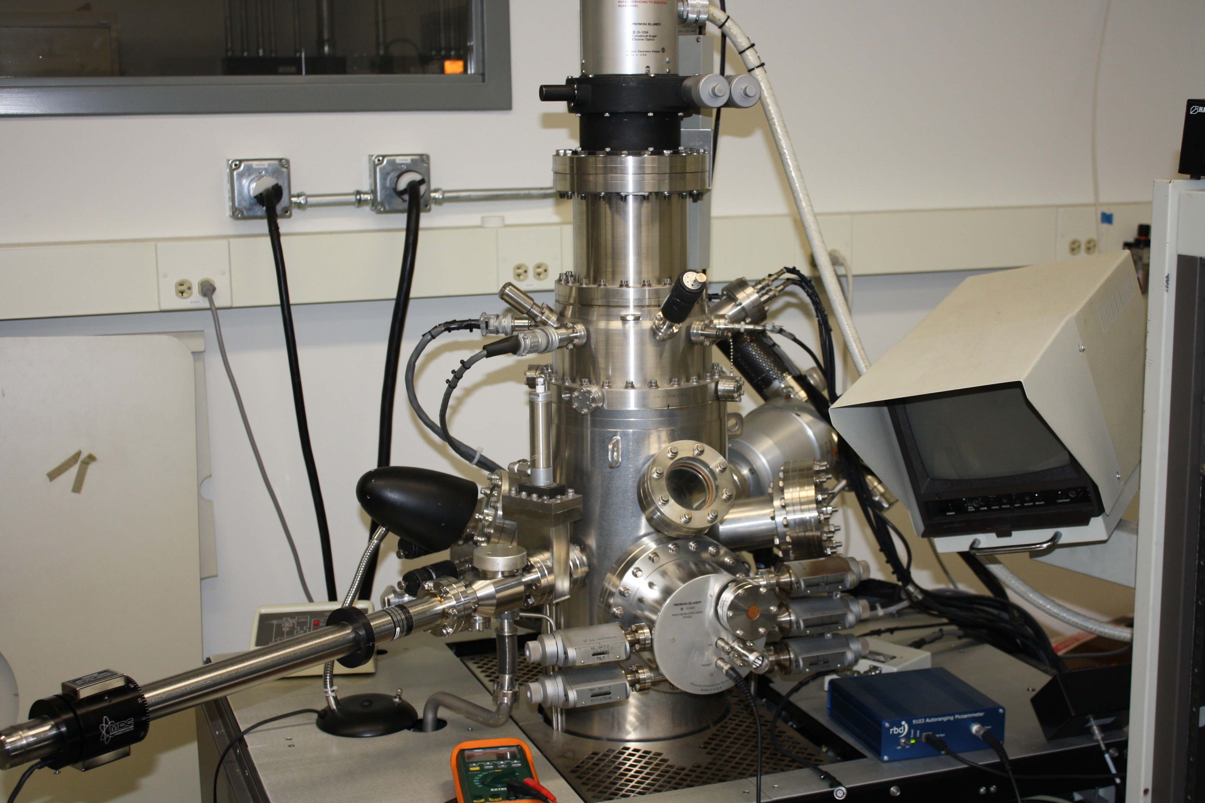 |
Our Auger electron spectrophotometer (AES) is used to study of surfaces of samples. This system has mapping and depth profiling capabilities to provide additional information about the sample uniformity. This system is a Perkin Elmer Phi 600. |
| Fourier Transform Infrared Spectroscopy (FTIR) | |
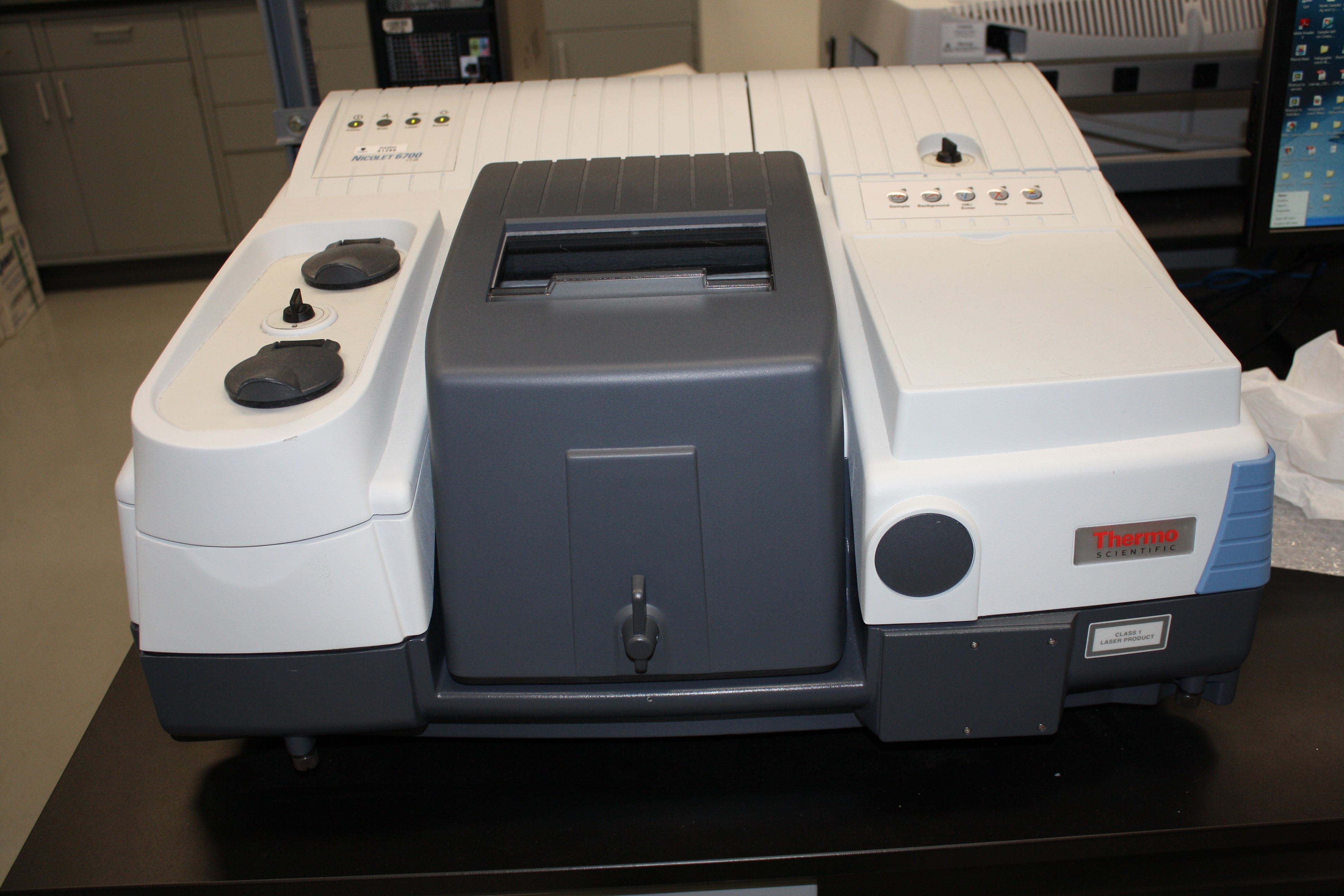 |
Our FTIR spectrometer completes IR spectroscopy over small areas. This system is purged with nitrogen to minimize the absorbance of water and oxygen in the sample chamber. Our system is capable of measuring in transmission and reflection mode. This a Thermo Scientific Nicolet system. |
| Inline SE | |
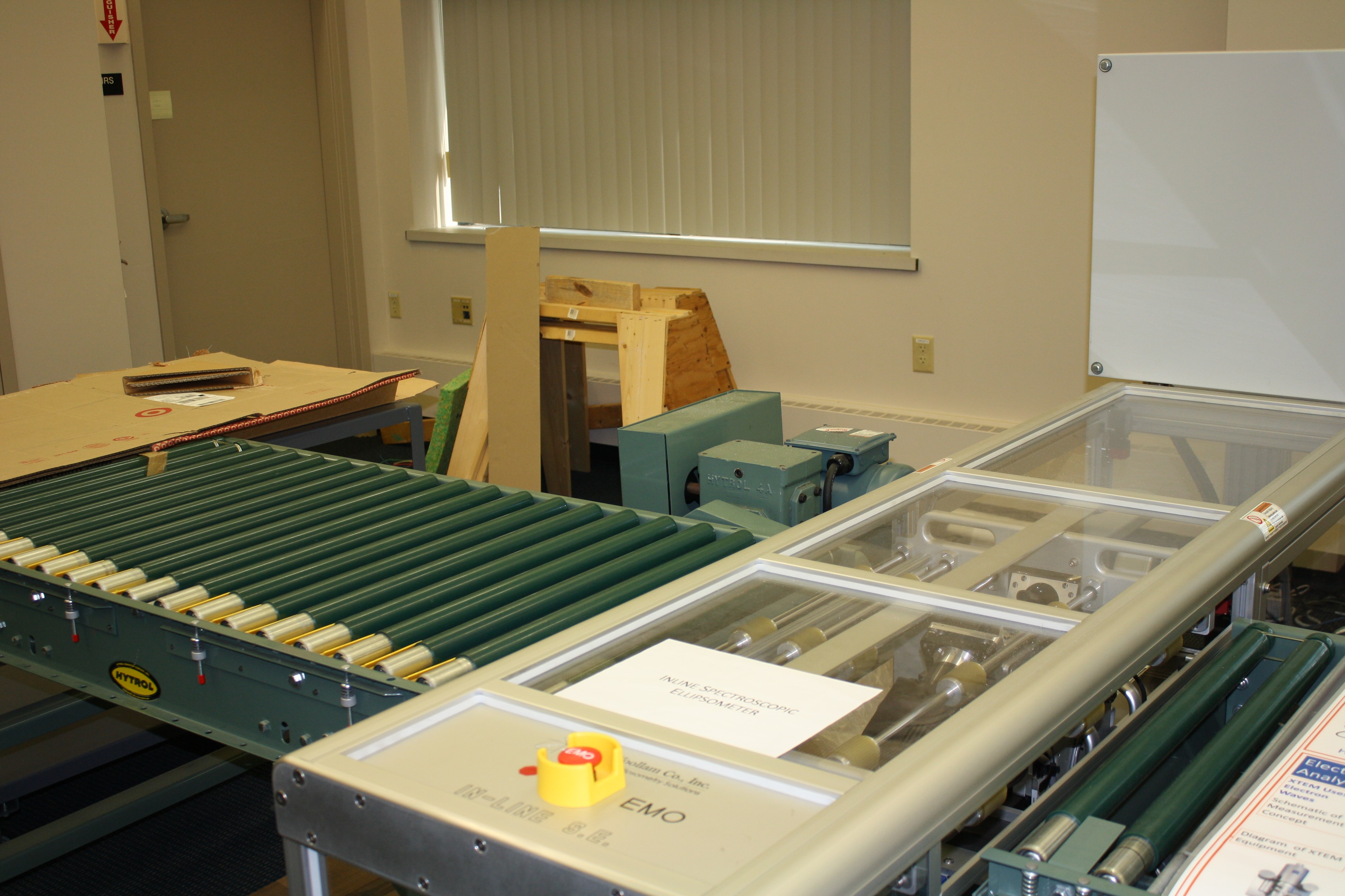 |
Our inline spectroscopic ellipsometer is a fixed angle rotating compensator ellipsometer integrated within a conveyor system designed for inline monitoring of photovoltaics (PV) panels. The motion of the PV panel is driven by a roller conveyor so that the panel passes a fixed optical monitoring station where the rotating-compensator multichannel ellipsometer performs the mapping function. This system was made by J.A. Woollam. |
| IR-VASE | |
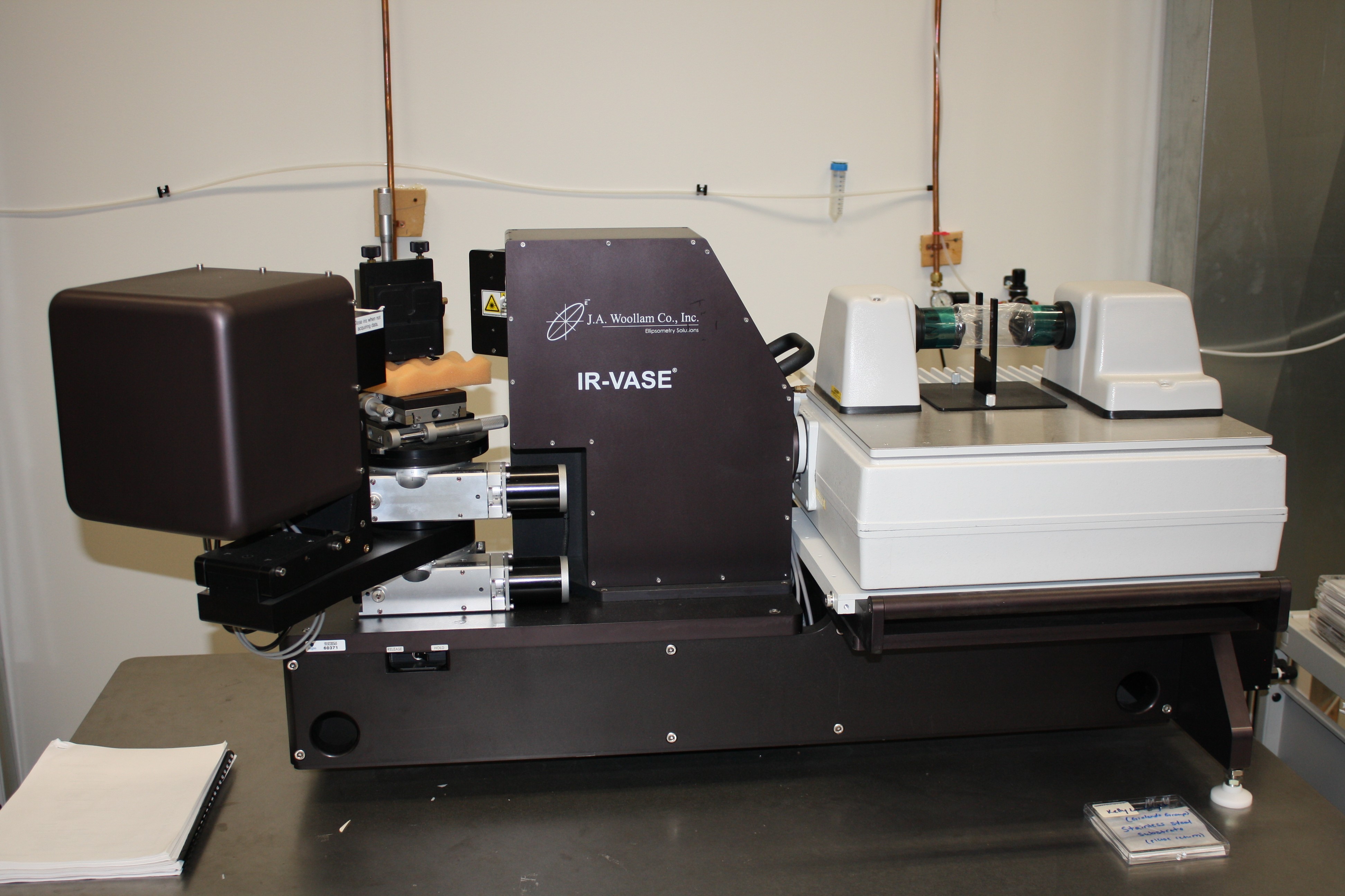 |
Our IR-VASE is a variable angle rotating compensator ellipsometer that covers a spectral range from 1.7 to 30 microns. It includes the features of Fourier Transform Infrared (FTIR) spectrometer as the light source. This system is a J.A. Woollam IR-VASE. |
| Large Area Mapping Ellipsometer (AccuMap) | |
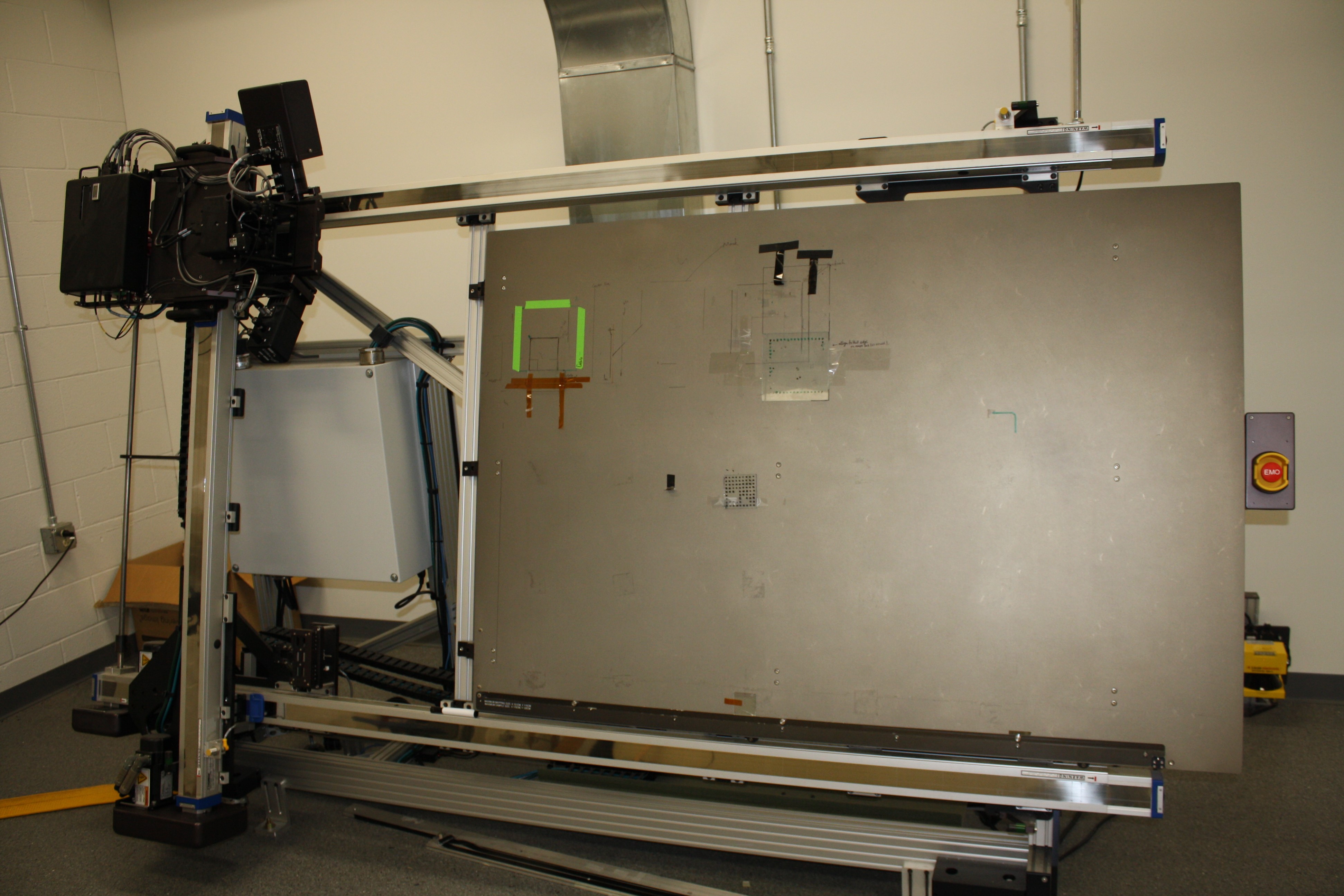 |
Our large area mapping ellipsometer is a fixed angle rotating compensator ellipsometer mounted on an x-y translation table used for large area mapping. This instrument is capable of scanning samples up to 1.5 m x 1.0 m size with a spatial resolution of ~ 1 millimeter in both directions. This system is a J.A. Woollam Accumap system. |
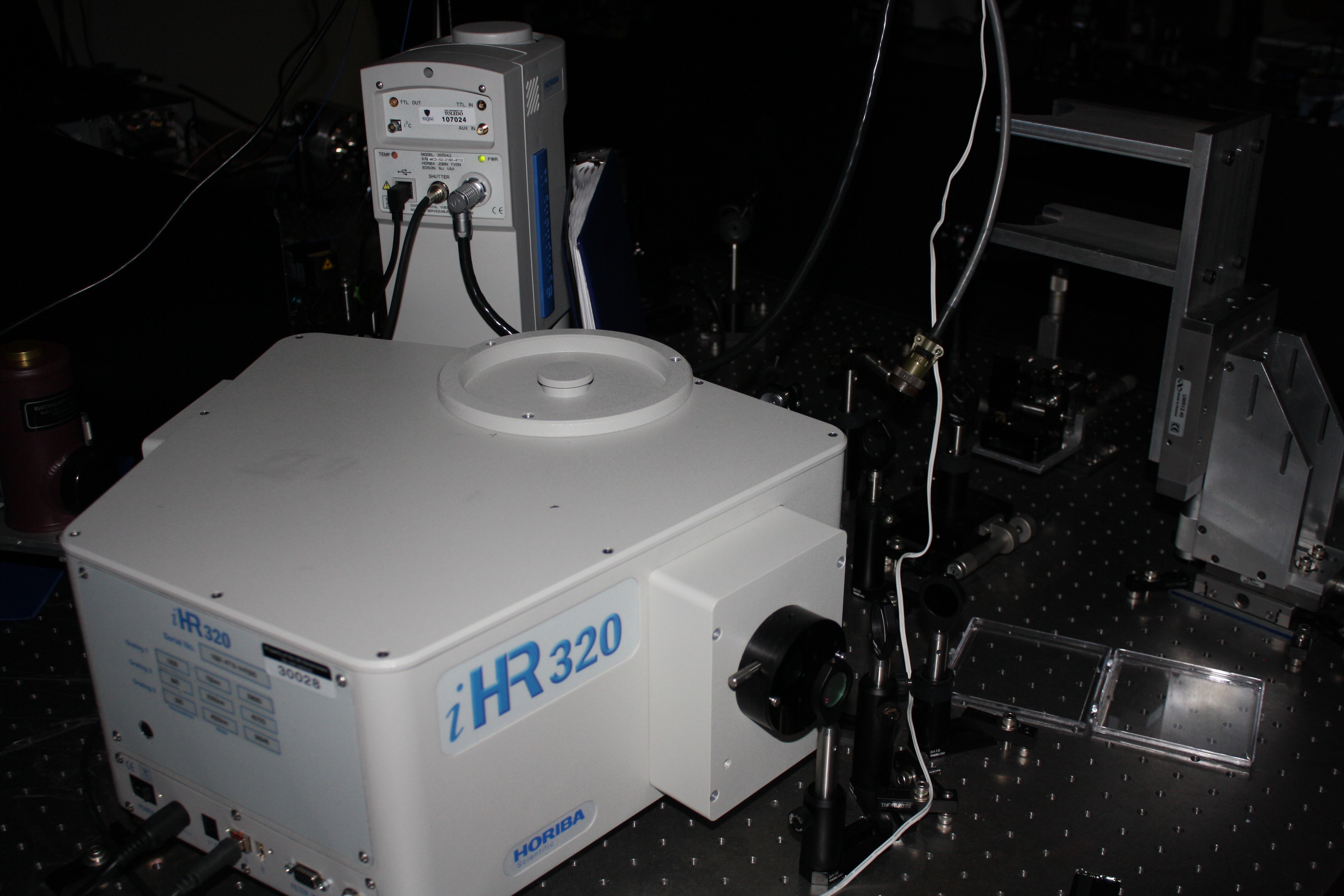
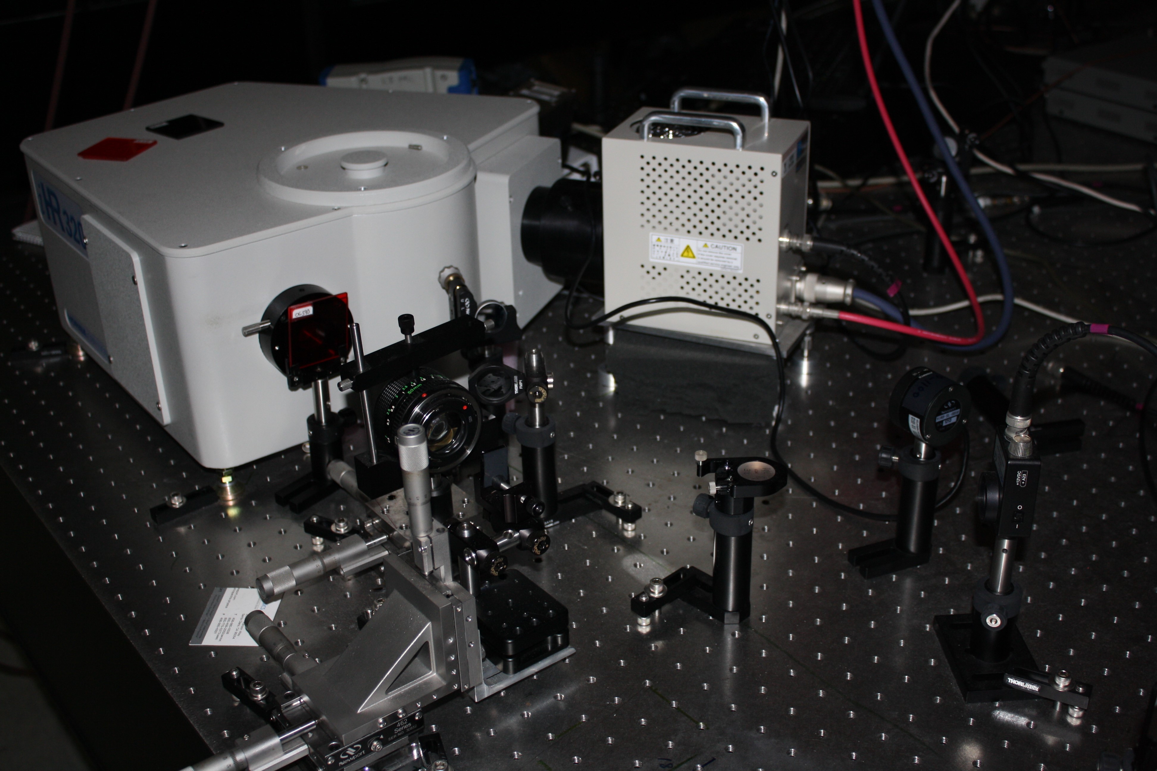
| Photoluminescence Excitation Mapping System | |
 |
Our photoluminescence excitation mapping system provides tunable excitation from 500 nm to 1000 nm and emission detection from 900 nm to 1700 nm. The instrument offers rapid, robust, and sensitive detection, and was used to perform PL mapping measurements. This system is a custom built system that attaches to a Thermo FT-Raman system. |
| Photoluminescence System | |
 |
Our photoluminescence system allows for characterization of semiconductor materials. The power of the excitation sources (405 nm, 532 nm, and 633 nm) are fully controllable. The system can detect PL emission from 400-1700 nm. |
| Photothermal Deflection Spectroscopy (PDS) | |
 |
Our photothermal deflection system operates over a spectral range of 1400 – 800 nm (0.89 – 1.55 eV) in 10 nm steps. The monochromatic pump beam is modulated with a mechanical chopper at 0.2 Hz. The probe beam is a 633 nm laser of nominally 300 μm cross sectional diameter. During PDS measurement, each film is immersed in a quartz cuvette containing a C6F14 fluid (Fluorinert FC-72, Synquest Laboratories). This system is a Sciencetech Inc. PTS-3-PTD. |
| Profilometer | |
 |
Our profilometer measures the vertical depth of a material across a horizontal length using a diamond stylus that transverses laterally across the sample. This equipment can be employed to measure etch depth, deposited film thickness, and surface roughness. This is a Veeco Dektak 150. |
| Raman Spectrophotometer | |
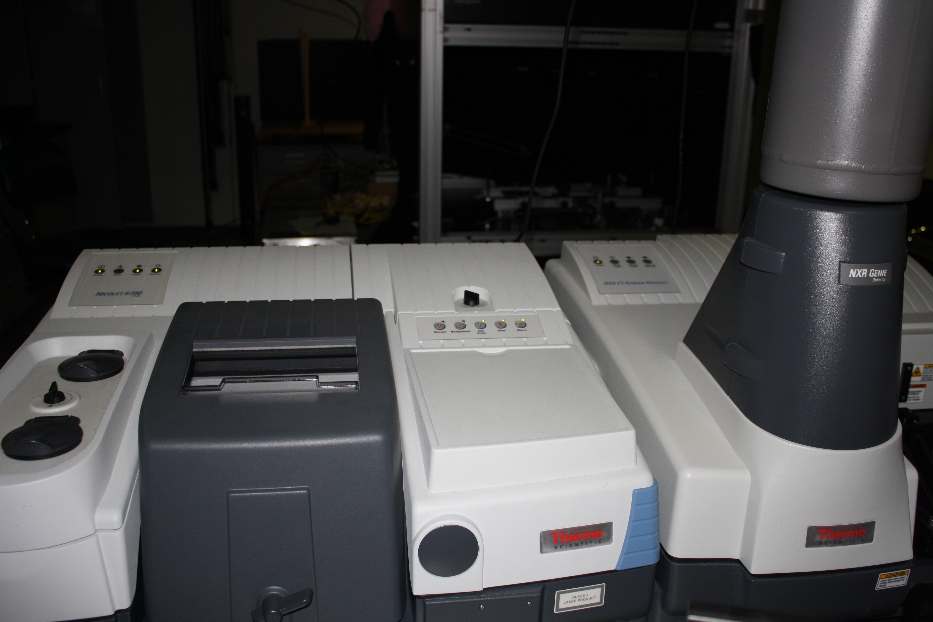 |
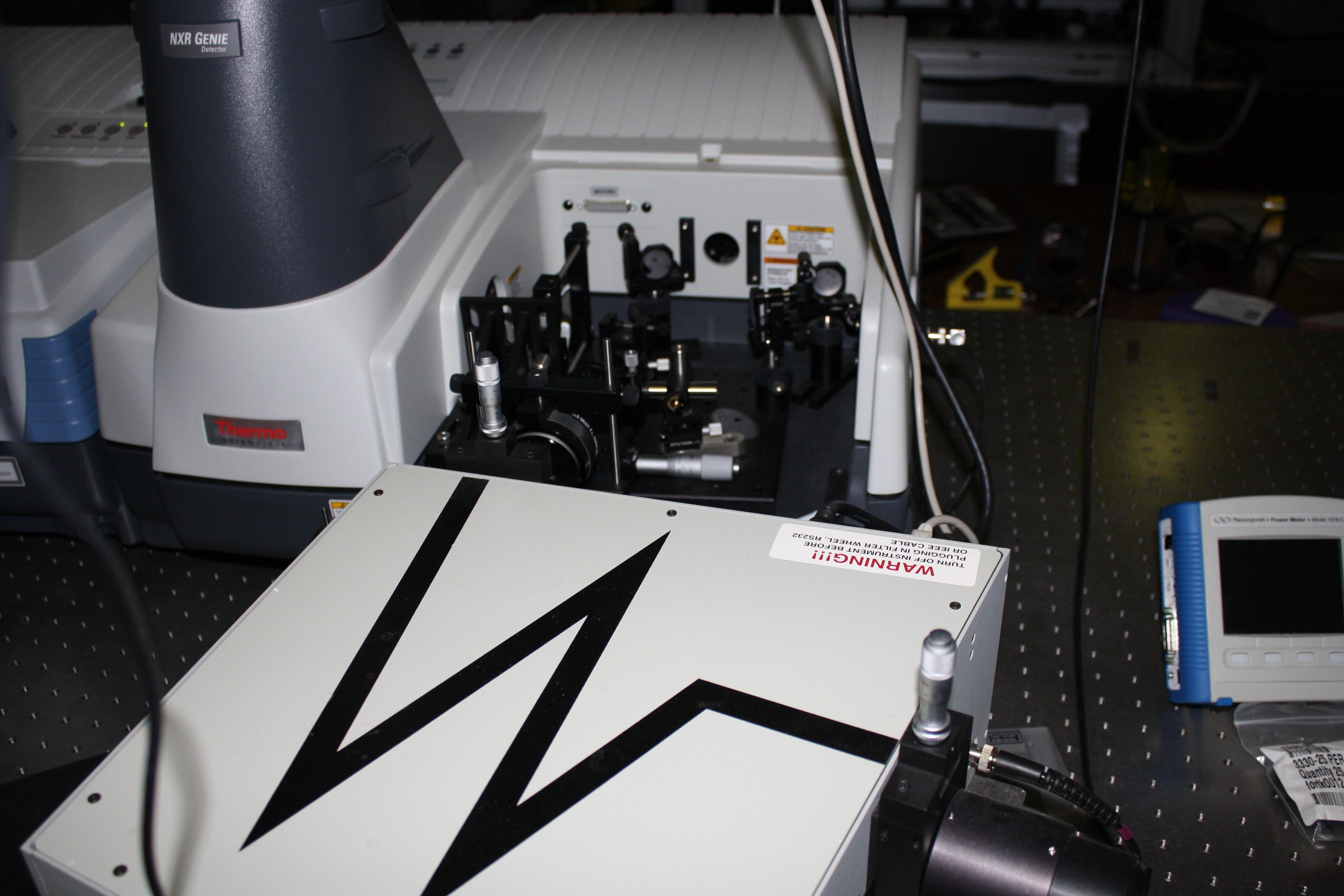
Our FT-Raman spectrometer is equipped with a 966 nm diode laser for excitation and a liquid nitrogen cooled germanium detector for measuring sample emission from 825 nm to 1700 nm. This system is a Thermo Scientific Nicolet NXR FT-Raman Spectrometer. |
| Sheet Resistance Measurement | |
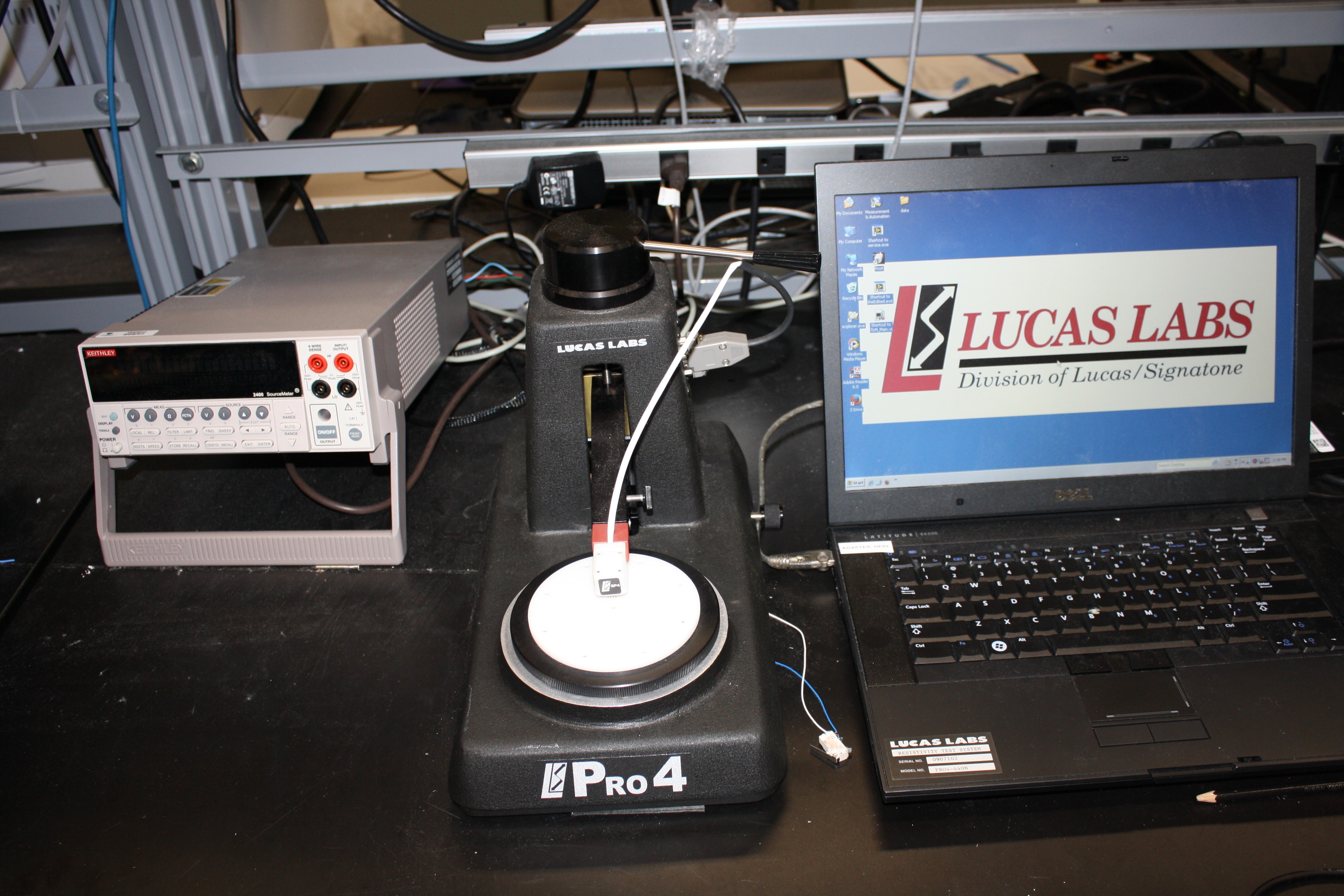 |
Our 4-point probe measures the sheet resistance of a thin film. We have multiple head configurations, each having pins with different sharpness. This system was made by Lucas Signatone. |
| Small Area Mapping Ellipsometer | |
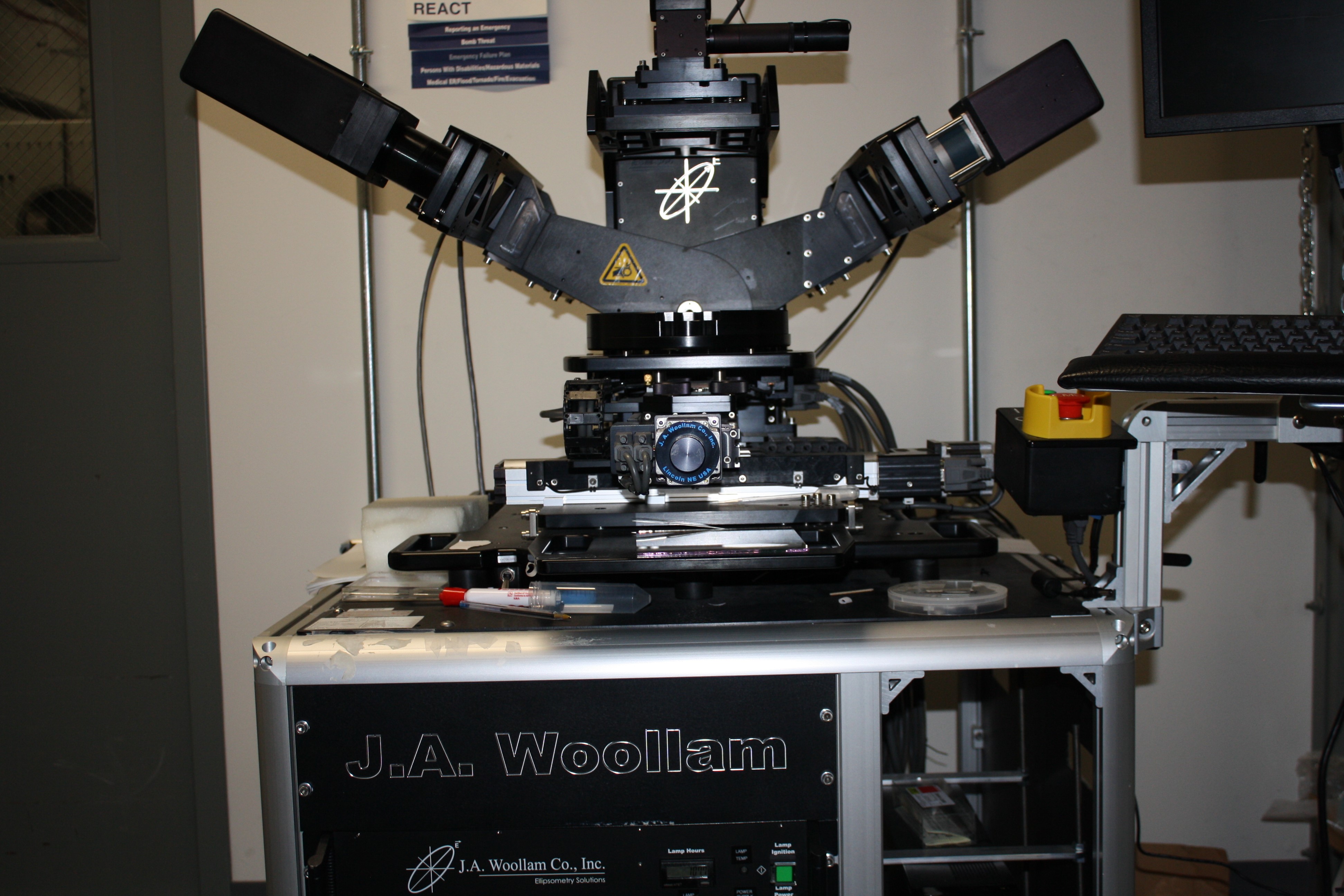 |
Our small area mapping ellipsometer is a variable angle rotating compensator ellipsometer mounted on an x-y translation table used for small area mapping. This instrument is capable of scanning samples up to 0.2 m x 0.2 m size with a spatial resolution of ~ 1 millimeter in both directions. This system was made by J.A. Woollam. |
| Spectrophotometer | |
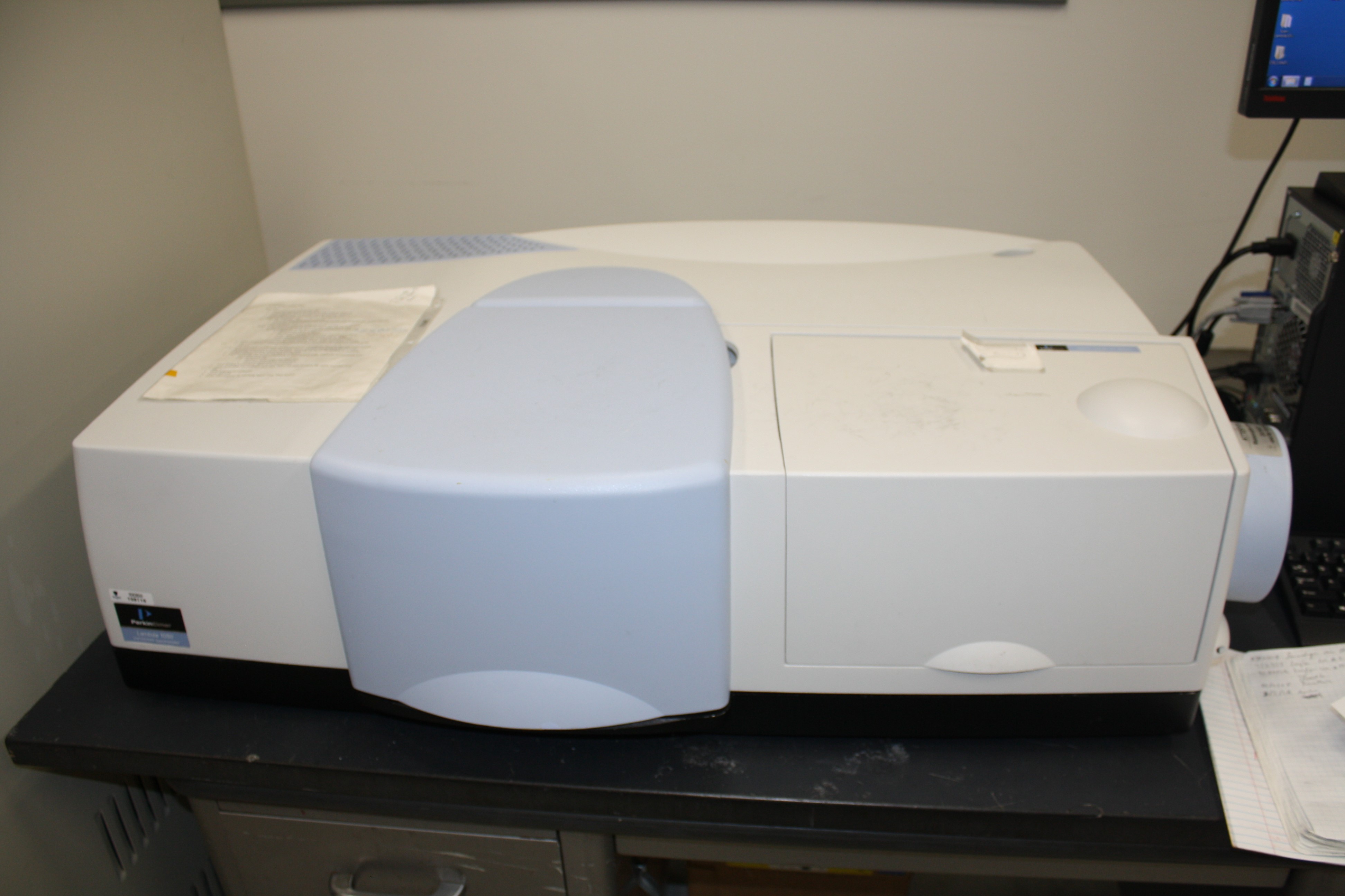 |
Our spectrophotometer measures the transmission and reflection from 190 nm to 3300 nm. In addition, we have multiple accessories that allow for variable angle and polarization measurements. This is a Perkin Elmer Lambda 1050 system. |
| Thermogravimetric Analysis System | |
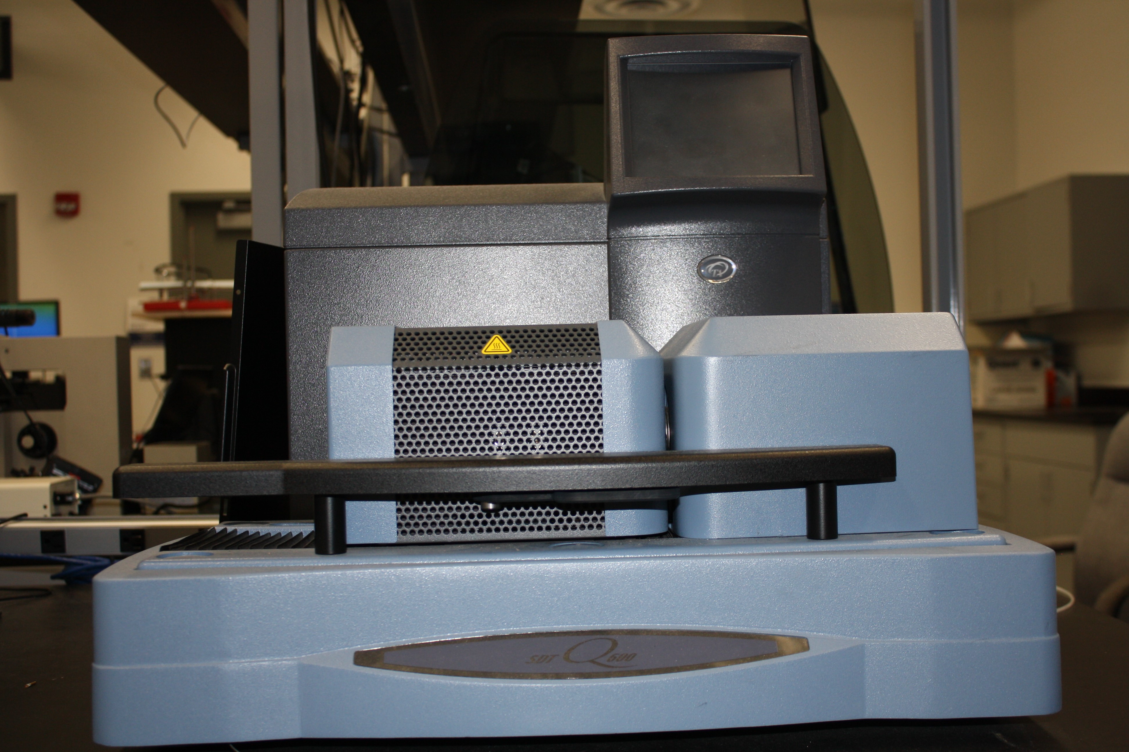 |
Our thermogravimetric analysis (TGA) and differential scanning calorimetry (DSC) system is capable of reaching 1300 degrees C on a small sample. The Pt boats allow for a wide range of gases to be used during heating. This is a TA Instruments Q600 system. |
| THz Ellipsometer | |
 |
Our terahertz ellipsometer is a variable angle rotating compensator ellipsometer capable of spectroscopic ellipsometry (SE) measurement in the ranges of 0.1–1.5 THz. This is a J.A. Woollam THz Ellipsometer. |
| Time-resolved Photoluminescence System | |
 |
Our time-resolved photoluminescence (TRPL), also referred to as Time Correlated Single Photon Counting (TCSPC), system utilizes an ultrafast laser pulse to excite the material while advanced electronics record the time delay between excitation and the detection of an emitted photon. This system has a wide range of excitation (450-1650 nm) and detection wavelengths (400-1700 nm). This system was built in-house. |
| Transient Absorption System | |
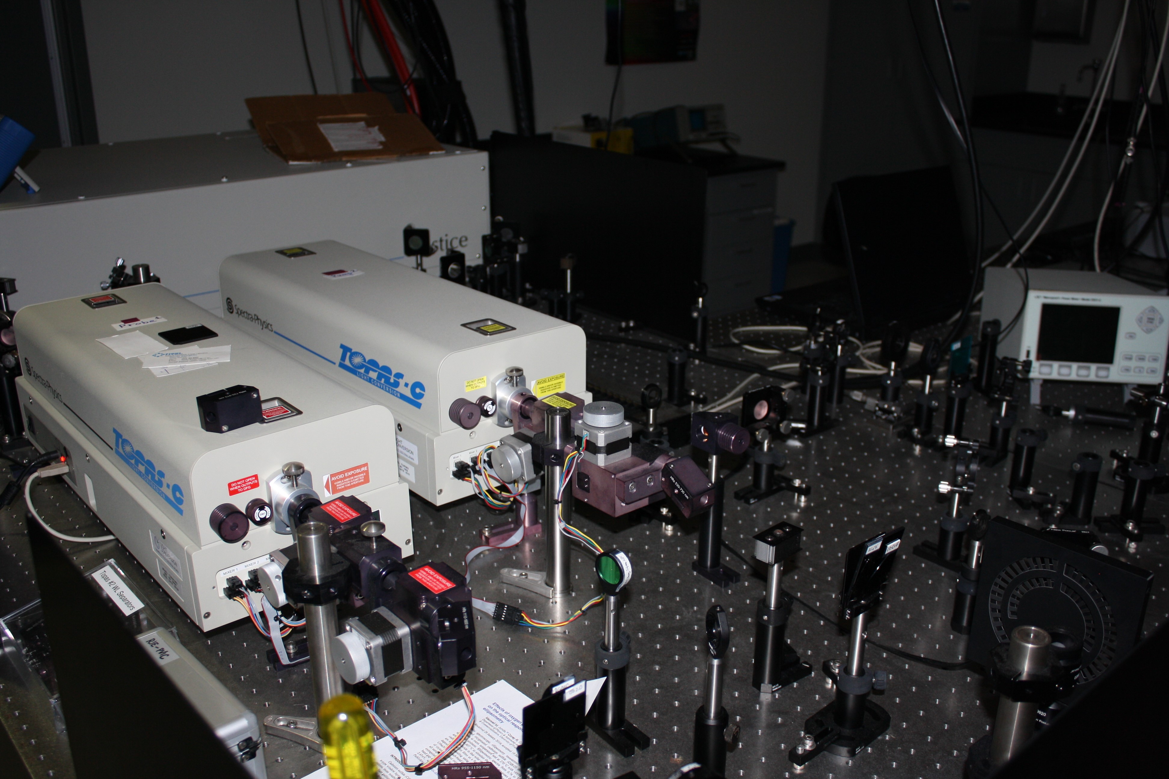 |
Our transient absorption (TA) system has the capabilities to pump from 270 nm to 3 μm and probe from 300 nm out to 9 μm independently from each other. The TA system consists of a Spectra Physics Solstice Laser system centered at 798 nm with ~100 fs laser pulse, repetition rate of 4 kHz, and pulse energy of 925 μj. |
| Transient Photocurrent Spectroscopy | |
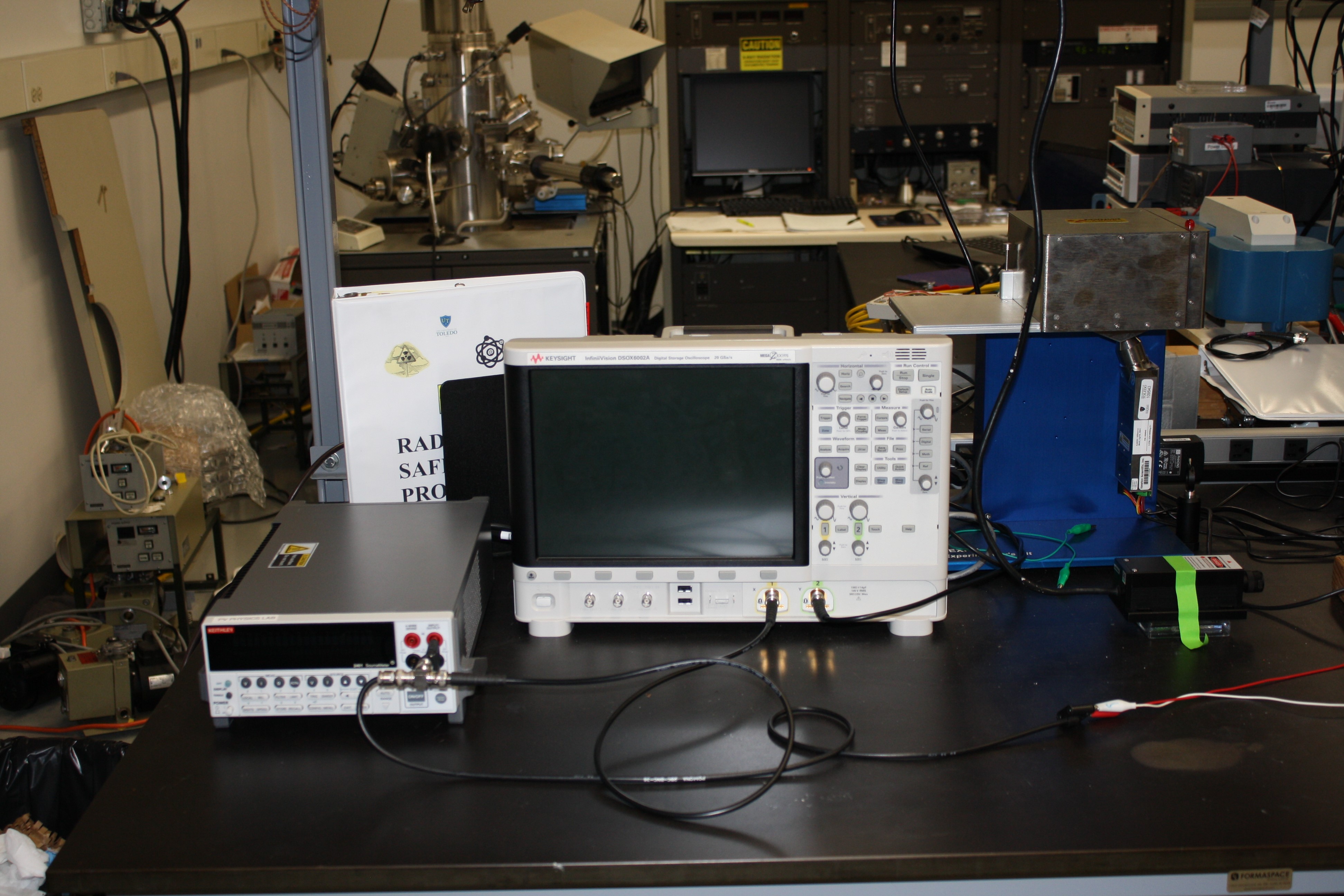 |
Our transient photocurrent spectroscoper is comprised of a Keysight InfiniiVision DSOX6002A ultrafast (20 GSa/s) digital oscilloscope, a Keithley 2400 source meter, a Keithley 6514 electrometer, a Newport low-noise current preamplifier (70710), a 473 nm laser, and a mini-X x-ray source. The system can be used to measure mobility in materials and response times of photodetectors. |
| Van der Pauw Hall Effect Measurement System | |
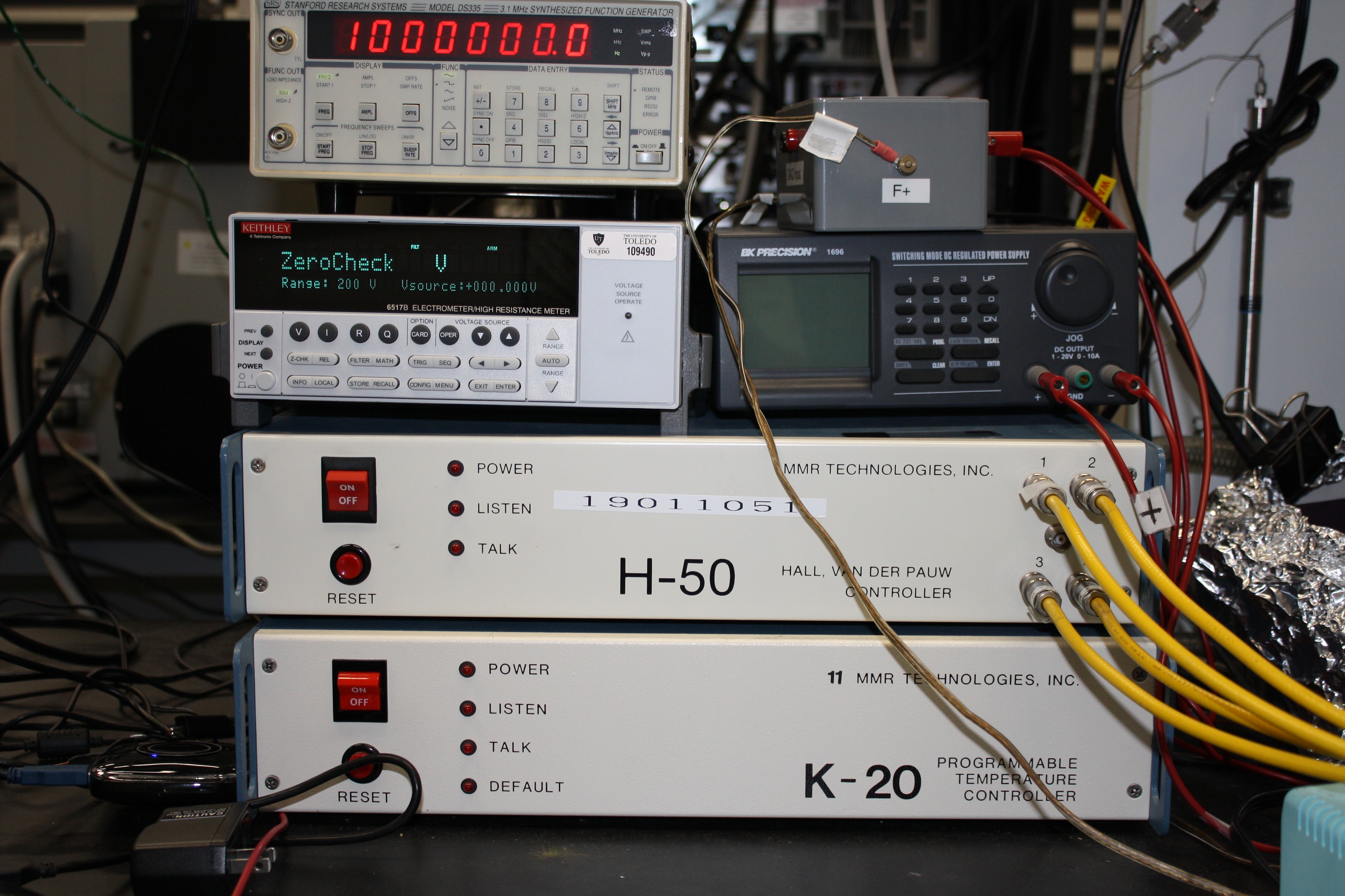 |
Our Hall effect measurement system uses the van der Pauw measurement technique to measure the sheet resistance, doping type, carrier density, and mobility of majority carrier. The variable temperature Hall Effect measurement with temperature ranging from 70 K to 500 K is feasible. |
| VUV-VASE | |
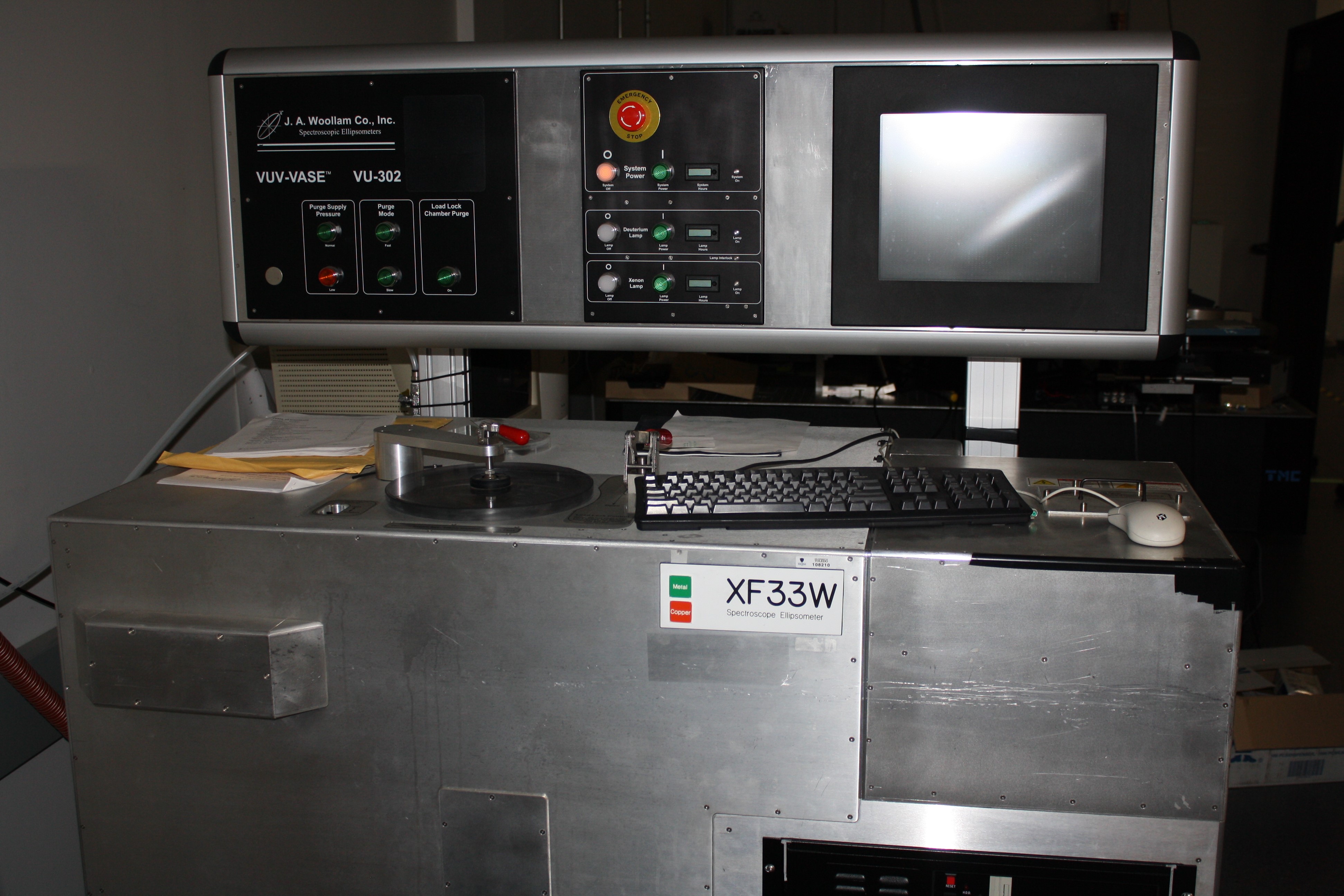 |
Our VUV-VASE is a variable angle rotating analyzer ellipsometer that allows measurements into the vacuum ultraviolet (down to 146 nm). Dual deuterium/xenon arc lamp light sources enable SE measurement in the range of 140 to 1700 nm. This is a J.A. Woollam VUV-VASE system. |
| V-VASE | |
 |
Our V-VASE is a variable angle rotating analyzer ellipsometer system operating in spectral range of 193-2500 nm. Features included are (i) vertical sample stage with vacuum mount can accommodate up to 200 mm diameter samples, (ii) automated angle of incidence from 15° to 90°, (iii) auto-retarder technology that provides highest accuracy on SE measurement, and (iv) measures depolarization, anisotropy, and Mueller-matrix data. This is a J.A. Woollam V-VASE system. |