

Micro CT and Skeletal Research Facility
Micro CT and Skeletal Research Resource Core, led by Dr. Piotr Czernik, assists researchers with comprehensive analysis of bone structure, imaging and quantification of lipids in bone marrow and soft tissue samples, in vivo assessment of body composition, bone mineral density and bone mineral content in small rodents. The facility is equipped with a Scanco μCT 35 x-ray scanner, an HP Integrity rx2660 workstation dedicated to image acquisition, processing, and analysis, and a GE Lunar PIXImus II dual-energy x-ray absorptiometry (DEXA) system.
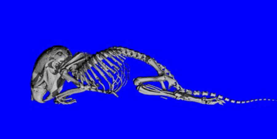
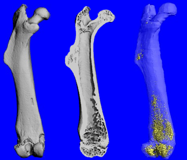
Volumetric imaging of bone and subsequent 3D image analysis allows for the quantitative assessment of a variety of structural properties including micro architecture, geometric parameters, volumes of individual compartments, distribution of bone mineral density, and alterations of bone structure induced by physical, pharmacological, or biological factors.
Standardized protocols for typical studies are listed below; customized procedures suited for unique projects can be developed upon request.
- Morphometric analysis of trabecular and cortical bone in rodents
- Morphometric analysis of human and large animal bones, cores and biopsies
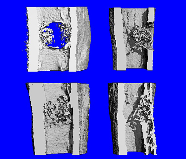
- Analysis of bone injury healing in animal models
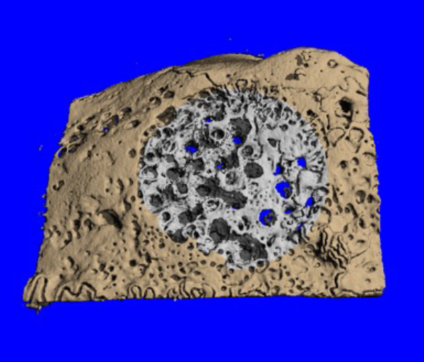
- Assessment of porosity in cortical bone
- Distribution and quantification of lipid content in bone marrow and soft tissue
- Computation of cross-sectional geometry of long bone
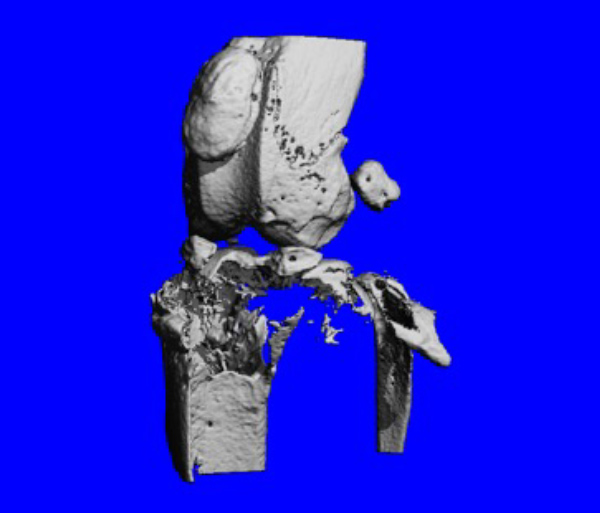
- Assessment of osseointegration and resorption of bone artificial implants and cements
- Preparation of publication quality images, videos, and STL surface models
The facility renders conventional and customized approaches to 3D imaging and analysis of a variety of specimens and provides assistance in experimental design.
Services
- Morphometry of human and animal bone
- Bone injury healing in animal models
- Bone porosity
- Lipid content in bone marrow
- Osseointegration of implants and cements
- 2D and 3D hi-res images, videos, and STL models
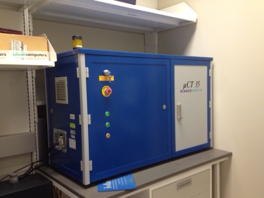
μCT 35 scanner
2009-2023
- Department of Bioengineering
- Department of Physiology and Pharmacology
- Department of Orthopaedic Surgery
- Department of Neurology
- Department of Cell and Cancer Biology
- Department of Dentistry
- Department of Neurosciences
- Ohio University
- Scripps Institute
- University of Kentucky
- Stryker Corporation
- Tissue Regeneration Systems, Inc.
- Vivorte, Inc.
- Dentsply International, Inc.
Bone marrow lipids
Spinal fusion

Implant integration
Bone injury healing
Cancer metastasis
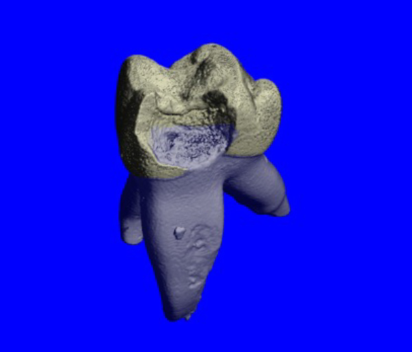
Tooth decay
Contact:
Peter Czernik, Ph.D.
419.383.4173
piotr.czernik@utoledo.edu