

Microscopy
Super Resolution Microscope
Nikon N-STORM Super Resolution Microscope & FRET
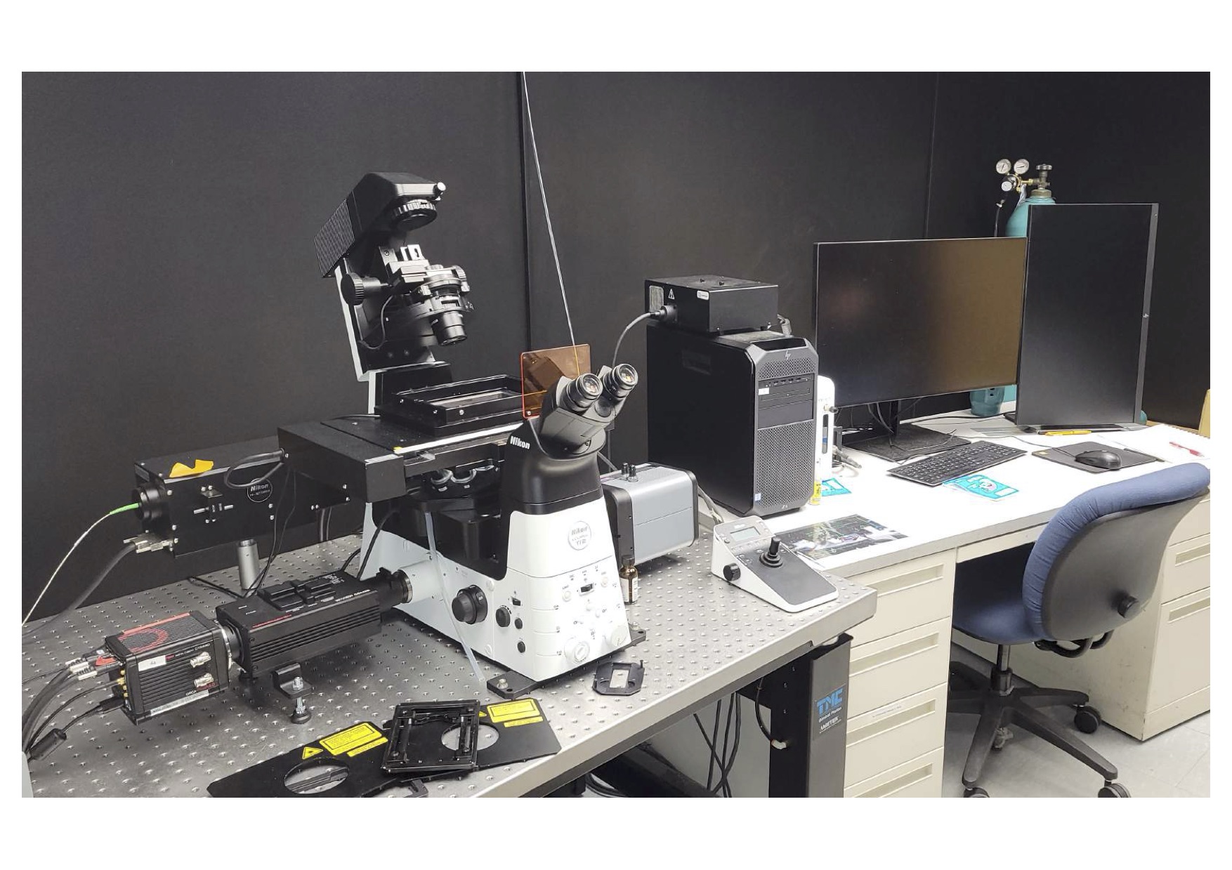
- Stochastic optical reconstruction microscopy
- Lateral resolution of ~20 nm
- Axial resolution of ~50 nm
- Multi-color imaging capability
- High definition, high density images
- NIS-Elements Software
- Multi-channel images
- Time lapse
- Multipoint functionality
- Image stitching
- Multidimensional image display
- 2D/3D Deconvolution
-
FRET (Fluorescence (Förster) Resonance Energy Transfer) capabilities
Contact: Chris Gianopoulos
Scanning Electron Microscopy (SEM)
JEOL JSM-7500F with EDS Detector Attachment
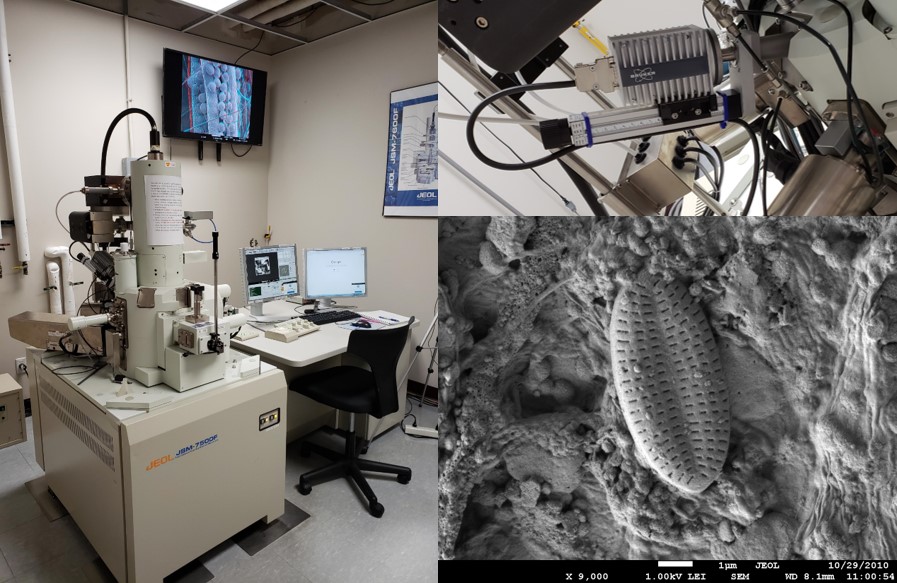
- Cold Cathode Field Emission Microscope
- Analyses samples up to dia. = 200 mm x height = 10 mm
- Various detectors, including:
- LABE - STEM - EBIC - EDS
- Cyber Enabled
- EDS attachment can be used for elemental mapping
- For an introduction to EDS theory and considerations for sample analysis see this video from the CMSC
- Gold and Carbon specimen coating available
- Typical Operations Manual can be found here
- A Guide to Scanning Microscope Observation can be found here
- Here is an informative video about choosing the right SEM parameters: https://youtu.be/eOyfoMRHfgE
- All samples must be completely dried before being placed in the SEM.
If you need help, please contact Dr. Britney Eggly
*Image above taken on our instrument of diatom
Stereoscopic Light Microscope
Olympus SZX7
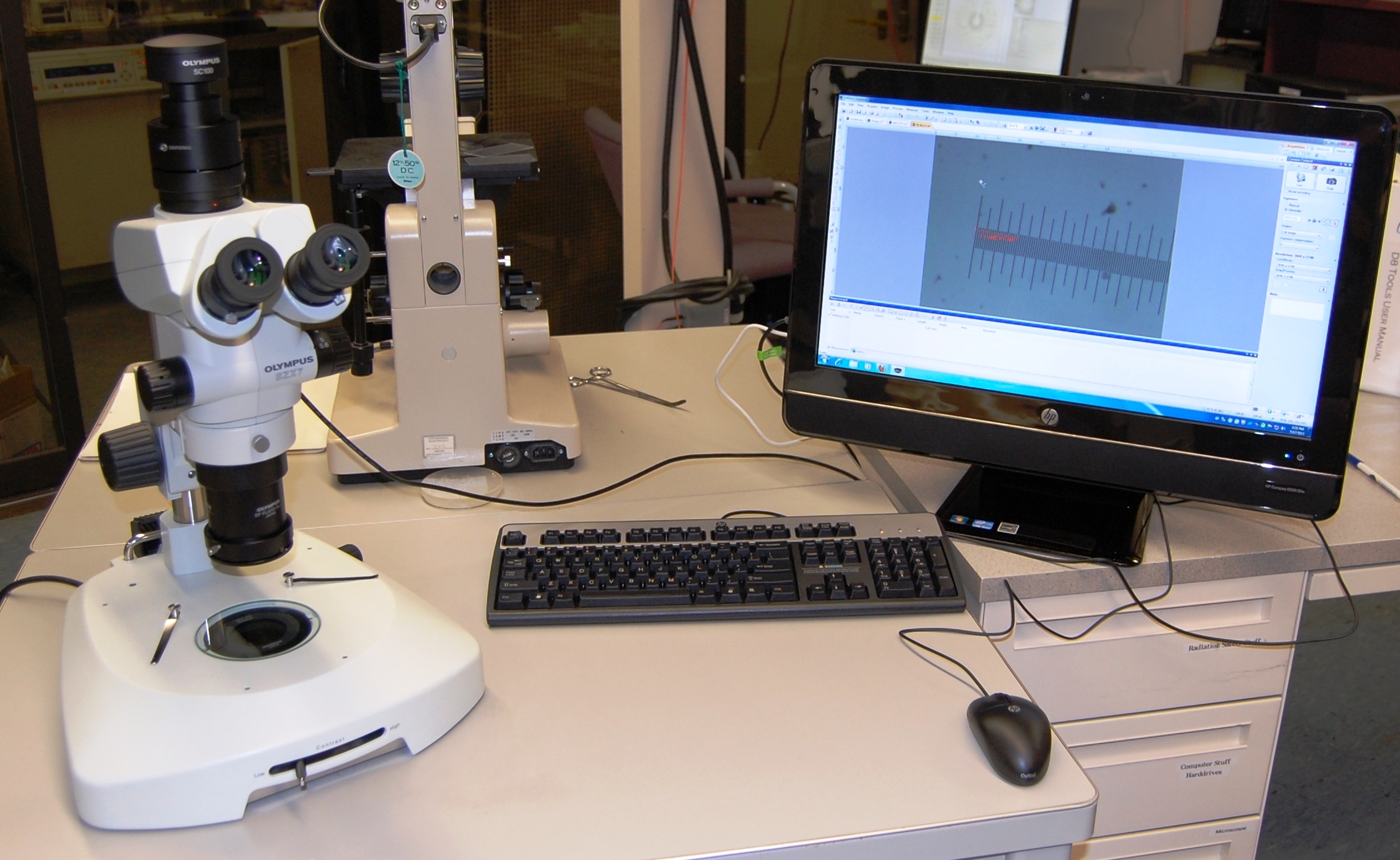
- Olympus SC100 camera directly attached
- Digital imaging
- Live video recording
- Images and measurements can be taken and viewed via a computer
Here are the manuals for the Microscope and Illuminator Stand and a short Video explaining the proper setup.
Contact: Dr. Kristin Kirschbaum